- *Corresponding Author:
- R. Jhorawat
Department of Nephrology, SMS Medical College and Hospital, J. L. N. Marg, Jaipur-302 004, India
E-mail: jhorawat2000@gmail.com
| Date of Submission | 04 December 2014 |
| Date of Revision | 13 November 2015 |
| Date of Acceptance | 08 February 2016 |
| Indian J Pharm Sci 2016;78(1):159-161 |
This is an open access article distributed under the terms of the Creative Commons Attribution-NonCommercial-ShareAlike 3.0 License, which allows others to remix, tweak, and build upon the work non-commercially, as long as the author is credited and the new creations are licensed under the identical terms.
Abstract
Nonsteroid antiinflammatory drugs have been implicated as nephrotoxic drugs, causing both acute and chronic adverse effects that range from reversible ischemia to chronic kidney disease and urothelial tumors to renal cell carcinoma specially papillary subtype. We report one case of collecting duct (Bellini duct) renal cell carcinoma in patient with analgesic-abuse nephropathy. This young individual was suffering from ankylosing spondylitis since the age of 16 years and was consuming diclofenac and paracetamol (acetaminophen) combination for >15 years. He developed hypertension, secondary glomerulopathy, chronic kidney disease and collecting duct renal cell carcinoma.
Keywords
NSAIDs, analgesic-abuse nephropathy, renal cell carcinoma, collecting duct renal cell carcinoma, clear cell renal cell carcinoma
Non-steroidal antiinflammatory drugs (NSAIDs) are one of the most commonly prescribed drugs, especially in chronic inflammatory disease. However, they are not without side effect that including acute and chronic, can range from reversible ischemia to chronic kidney disease and urothelial tumors to renal cell carcinoma (RCC). The NSAIDs has been implicated in the causation of the papillary subtype of RCC [1].
Collecting duct (Bellini duct) renal cell carcinoma (CDRCC) occurring in 0.4-2.0% of cases of renal cell carcinoma (RCC) which make us to depend on case report or case series for our knowledge to this rare subtype. Till now we have indirect evidence of relationship of NSAIDs in the causation of CDRCC. This is perhaps the first case, in which NSAIDs are directly related in the causation of this rare subtype.
This was a case of thirty eight years old young male who was symptomatic for last twenty two years with low back pain and bilateral pain and swelling in ankle joints with early morning stiffness. No pain and swelling in other joints. He consulted a physician and was started on pain killer containing diclofenac and paracetamol (acetaminophen) combination, which relieved his pain significantly. Thereafter, he used to take the same medication whenever he feels increase or worsening of his back pain. He was not on regular follow up to any physician and continue to consume this medication for >15 years.
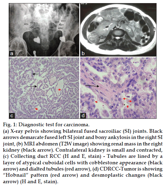
Now, after twenty years, he was admitted with symptoms of generalized body swelling and headache. He was found to be hypertensive and excreting proteins in his urine in nephrotic range (24 h urine protein=4944 g total volume=1600 ml). The abdomen sonography was showing mass in his right kidney (lower pole) and contracted `ral kidney´. His renal function test was deranged (serum creatinine=5.7 mg/dl, serum urea=157 mg/dl and was anemic (hemoglobin=7.8 gm/dl). Other investigation was [Na+]=140 mEq/l, [K+]=5.3 mEq/l, serum albumin=3.0 g/dl, serum total protein=5.8 g/dl, serum alkaline phosphate=89 IU/l, serum cholesterol=231 mg/dl. He was evaluated for his basic disease. X-ray pelvis was showing bilateral fused sacroiliac joints (fig. 1a). MRI abdomen confirmed solid mass in the right kidney (fig. 1b). HLA-B27 was positive and rheumatoid factor was negative with raise ESR and positive CRP (qualitative). The diagnosis of ankylosing spondylitis with analgesic-abuse nephropathy (secondary FSGS) and incidental detected renal mass? RCC was made. The right side nephrectomy was done with histopathology of the mass was showing collecting duct type RCC (CDRCC), as shown in fig. 1c and d. He remains dialysis dependent during follow up. He was on regular hemodialysis for one and a half month; however, he demised after 2 months.
Figure 1: Diagnostic test for carcinoma.
(a) X-ray pelvis showing bilateral fused sacroiliac (SI) joints. Black arrows demarcate fused left SI joint and bony ankylosis in the right SI joint, (b) MRI abdomen (T2W image) showing renal mass in the right kidney (black arrow). Contralateral kidney is small and contracted, (c) Collecting duct RCC (H and E, stain) - Tubules are lined by a layer of atypical cuboidal cells with cobblestone appearance (black arrow) and dialted tubules (red arrow), (d) CDRCC-Tumor is showing “Hobnail” pattern (red arrow) and desmoplastic changes (black arrow) (H and E, stain).
As we know NSAIDs are drugs with ‘two-edge sword’. Use of certain analgesics, including aspirin and non-aspirin NSAIDs have been associated with reduced risk of breast, prostate, and colorectal cancers. On the other hand, they increase the risk of urinary tract carcinoma and RCC. Recently, a comprehensive meta-analysis of studies dedicated to the relationship between the three most commonly used analgesics (acetaminophen, aspirin and non-aspirin NSAID) and kidney cancer risk, had shown that acetaminophen and non-aspirin NSAIDs increased the risk of RCC [1]. In our case also, he was consuming paracetamol (acetaminophen) and diclofenac for more than a decade which might predispose him for RCC.
NSAIDs are reported to increase both RCC and uroepithelial tumors. Rocha et al. in their study on rat had shown that aspirin, salicylic acid and acetaminophen reduced number of rapidly proliferating cell by 50% in inner medullary collecting duct [2]. Acetaminophen, in addition, arrests most cell in late G1 and S phase [3]. It produced mixed form of cell death with both oncosis (swollen cells and nuclei) and apoptosis. Acetaminophen inhibits DNA synthesis and cause chromosomal aberration due to inhibition of ribonucleotide reductase. These genotoxic effects have been postulated in carcinogenesis due to NSAIDs [2].
CDRCC are distinct tumors from clear cell RCC (CCRCC) which is a most common renal tumor. CDRCC develops from the collecting ducts in the renal medullary pyramid, whereas CCRCC arise from the convoluted tubules. There are no specific CT findings to distinguish CDRCC from CCRCC although medullary location, weak and heterogeneous enhancement, renal sinus involvement, infiltrative growth with preserved renal contours and a cystic component are frequently seen. Tumor cell in CDRCC show positivity with antibodies to Ulex European agglutinin 1 lectin, peanut agglutinin (PNA), vimentin, lysozyme, distal tubular markers (e.g EMA and high molecular weight cytokeratin) and negative for proximal tubular markers [4]. These markers, however, are not tested in our case.
In the available literature, CDRCC is indirectly related to NSAIDs. There is a case report, which highlighted coexistence of two subtype of RCC in the same individual at the same time i.e CDRCC and papillary RCC [4]. Another case report, which mention the occurrence of CDRCC subtype simultaneously with transitional cell carcinoma (TCC) in a same individual at the same time [5]. Both these tumors which were co-existent with CDRCC had been reported with NSAIDs as their risk factor. In addition, CDRCC shares some histological similarities and site of origin with papillary subtype of RCC and some common genetic abnormalities with TCC. Ours is the first case in which NSAID exposure directly reported to be associated with CDRCC. As CDCC has a very low incidence of all renal tumors, as a result, our knowledge of this tumor is limited.
In conclusion, CDRCC is a rare and aggressive subtype of RCC. Our available knowledge for this tumor arises from small case series and case reports. And our case directly relates that long exposure of acetaminophen and NSAIDs might be a risk factor for CDRCC also.
Financial support and sponsorship
Nil.
Conflicts of interest
There are no conflicts of interest.
References
- Choueiri TK, Je Y, Cho E. Analgesic use and the risk of kidney cancer: A meta-analysis of epidemiologic studies. Int J Cancer 2014;134:384-96.
- Rocha GM, Michea LF, Peters EM, Kirby M, Xu Y, Ferguson DR, et al. Direct toxicity of nonsteroidalantiinflammatory drugs for renalmedullary cells. ProcNatlAcadSci U S A 2001;98:5317-22.
- Hongslo JK, Bjørge C, Schwarze PE, Brøgger A, Mann G, Thelander L, et al.Paracetamol inhibits replicative DNA synthesis and induces sister chromatid exchange and chromosomal aberrations by inhibition of ribonucleotidereductase. Mutagenesis 1990;5:475-80.
- Matei DV, Rocco B, Varela R, Verweij F, Scardino E, Renne G, et al. Synchronous collecting duct carcinoma and papillary renal cellcarcinoma: A case report and review of the literature. Anticancer Res 2005;25:579-86.
- Pereira Arias JG, AtecaDíaz-Obregón R, Gutiérrez Díez JM, Ramírez Rodríguez MM, EtxezarrazgaZuluaga MC, BerreteagaGallastegui JR. Synchronous association of Bellini collecting ducts and pyelic transitional cell carcinoma.ActasUrolEsp 1997;21:990-6.
