- *Corresponding Author:
- P. C. Ghosh
Department of Biochemistry, University of Delhi South Campus, Benito Juarez Road, New Delhi-110 021, India
E-mail: pcghose@gmail.com
| Date of Submission | 20 April 2012 |
| Date of Revision | 03 January 2013 |
| Date of Acceptance | 05 January 2013 |
| Indian J Pharm Sci 2013;75(1):16-22 |
Abstract
The monensin, known to enhance the cytotoxicity of ricin and ricin-based immunotoxins is a very hydrophobic molecule and this limits its administration in optimum doses under in vivo conditions. In order to realise its full potential, monensin was intercalated into various liposomal formulations and its ability to potentiate the cytotoxicity of ricin liposomes in human epidermoid carcinoma (KB) cells was studied. It was observed that ricin cytotoxicity enhancing ability of monensin liposome depends on the surface charge as well as density and chain length of distearoyl phosphatidylethanolamine-methoxy polyethylene glycol present on the surface of liposomal monensin. Maximum potentiation on the cytotoxicity of liposomal ricin was observed by monensin entrapped in neutral liposome (106.5 fold) followed by negatively charged (94.2 fold) and positively charged liposome (90 fold). Studies on the effect of variation of density and chain length of distearoyl phosphatidylethanolamine-methoxy polyethylene glycol showed that neutral monensin liposomes having 2.5 mol% distearoyl phosphatidylethanolamine-methoxy polyethylene glycol with chain length of 2000 exhibits maximum potentiation (117.6 fold) on the cytotoxicity of ricin liposomes when the cellular uptake of monensin liposome was maximum (42.0%) and the zeta potential value on the surface of liposomes was −0.645. The present study has clearly shown that liposomal monensin is very effective in enhancing the cytotoxicity of liposomal ricin in human cancer cells and liposome can be used as in vivo deliver vehicle for monensin to potentiate the cytotoxicity of liposomal ricin to eliminate cancer cells.
Keywords
Human cancer cells, liposomal monensin, liposomal ricin, sterically stabilized liposomes, zeta potential
Ricin, a heterodimeric toxic protein (consisting of A‑ and B‑chain) is known to inhibit the synthesis of proteins in eukaryotic cells by cleaving a specific adenine residue from 28S rRNA presumably mediated by the N‑glycosidase activity of the A‑chain [1]. However, the potential use of ricin as an anticancer agent arising from its ability to arrest protein synthesis in eukaryotic cells is limited due to the nonselectivity of the B‑chain. Liposome has been used as delivery vehicle for delivery of ricin to cancer cells, but cytotoxicity of ricin reduced markedly following encapsulation into liposomes [2‑4]. It has been reported that free monensin significantly enhances the cytotoxicity of ricin encapsulated in liposomes in Chinese hamster ovary (CHO pro−) cells [4]. The enhancement of the cytotoxicity of liposomal ricin by free monensin was found to be significantly dependent on the lipid composition, charge and hydrophilicity on the surface of liposomes employed for the delivery of ricin [3]. However, monensin is a very hydrophobic molecule and this limits its administration in optimum doses under in vivo conditions in order to realise its full potential in the enhancement of toxicity of liposomal ricin. The development of a delivery system for this ionophore is, therefore, vital for exploitation of its role as potentiating agent under in vivo condition. Vasandani et al., for the first time reported that liposomes can be used as a delivery vehicle for monensin [5]. It has also been shown that liposomal monensin is very effective in enhancing the cytotoxicity of ricin in free form in mouse macrophage tumour cells [6] and under in vivo condition [5,7]. Subsequently, it has been reported by a number of investigators that liposomal monensin is very effective in enhancing the cytotoxicity of tumour cell specific immunotoxins for selective elimination of tumour cells [8‑10]. However, the relatively short half‑life of these liposomes was a major drawback for their limited use in vivo. To overcome this problem, new generations of liposomes, which have prolonged circulation time in the blood, have been developed. These liposomes are called stealth or sterically stabilised liposomes and are very stable in the circulation for prolonged period of time. It has been reported that the sterically stabilised liposomes of 70‑200 nm size containing 5‑7.5 mol% of distearoyl phosphatidylethanolamine-methoxy polyethylene glycol with chain length of 2000 (DSPE-mPEG-2000) exhibit maximum circulatory life [11]. It has been also reported that the surface modification of the liposome with PEG‑lipids of varying molecular weights of PEG (chain length) has prolonged circulation time in the blood by avoiding the uptake by liver and spleen [11‑13]. It has been reported by a number of investigators that the interaction of these liposomes with macrophages [12] and cancer cells [14,15] significantly influenced by the chain length of PEG. However, no work has been carried out so far to explore the efficacy of liposomal monensin on the enhancement of cytotoxicity of liposomal ricin. Therefore, attempts have been made during this investigation to intercalate monensin into various formulations of liposomes. The efficacy of monensin liposomes having different density and chain lengths of DSPE‑mPEG on the enhancement of fold potentiation of the cytotoxicity of liposomal ricin was studied in human epidermoid carcinoma (KB) cells. For this study, liposomes composed of soya phosphatidylcholine (SPC), cholesterol, phosphatidic acid (PA) and 5 mol% DSPE mPEG‑2000 are used for delivery of ricin, because the maximum enhancement of cytotoxicity of ricin by free monensin was observed in KB cells when ricin was delivered through this liposomal formulation [2].
Materials and Methods
Cholesterol, L‑a‑PA, stearylamine (SA), lactoperoxidase, monensin, 2,5‑diphenyl oxazole, 1,4‑bis [2‑(5‑phenyl oxazole)] benzene, Guar gum, penicillin‑G (potassium salt), streptomycin sulphate, 2.5% trypsin in HBSS (Hank’s balanced salt solution) and HEPES ((4-(2-hydroxyethyl)-1-piperazineethanesulfonic acid) were purchased from Sigma Chemical Co., St. Louis, USA. SPC was obtained as gift from Life‑care Innovations Pvt. Ltd., Haryana, India. DSPE‑mPEG (1000, 2000, 3000 and 5000) (distearoyl phosphatidylethanolamine polyethylene glycol) was purchased from Avanti Polar Lipids, Inc., Alabama, USA. Foetal calf serum (FCS) was obtained from Biological Industries Ltd., Beit‑Haemek, Israel. Sepharose CL 6B, Sephacryl S‑300 and Sephadex G‑25 were obtained from Pharmacia Fine Chemicals, Uppsala, Sweden. Powdered Roswell Park Memorial Institute (RPMI) 1640 (deficient) medium was obtained from Sigma Chemical Co., St. Louis, USA. Dulbecco’s Modified Eagle’s Medium (DMEM) purchased from Invitrogen Corporation, New York, USA. [3H] Leucine and Na125I were bought from American Radiochemicals Inc., St. Louis, USA.
Human KB cells were procured from National Centre for Cell Science, Pune, India and these cells were maintained in DMEM. The culture media were supplemented with 10% FCS, HEPES, NaHCO3, penicillin (100 units/ml) and streptomycin (100 mg/ml). Cells were then grown in a humidified incubator at 37°, 5% CO2 and 95% air atmosphere.
Purification and radioiodination of ricin
Ricin was purified from the seeds of Ricinus communis and radiolabelled with Na125I by lactoperoxidase according to the method reported earlier [16].
Liposomal entrapment of ricin
Liposomes composed of SPC, cholesterol, PA and DSPE‑mPEG‑2000 in a molar ratio of 40:45:10:5 were prepared by hand shaken method as described earlier [2]. Liposome size and zeta potential were determined using a Zeatasizer Nano ZS (ZEN 3600, Malvern Instruments, Worcestershire, UK) as presented in Table 1.
| Liposomal formulations for monensin | Molar ratio | Size (nm) | Zeta potential (mV) | *Intercalation efficiency (%) |
|---|---|---|---|---|
| SPC:chol:monensin (neutral) | 63:27:10 | 124 ± 26 | -4.256 | 54.2 ± 8.52 |
| SPC:chol:monensin:PEG-2000 | 62:27:10:1 | 142 ± 41 | -2.234 | 56.3 ± 5.48 |
| SPC:chol:monensin:PEG-2000 | 60.5:27:10:2.5 | 128 ± 43 | -0.645 | 54.1 ± 6.24 |
| SPC:chol:monensin:PEG-2000 | 58:27:10:5 | 112 ± 32 | -0.324 | 58.2 ± 3.48 |
| SPC:chol:monensin:PEG-2000 | 55.5:27:10:7.5 | 108 ± 24 | -0.233 | 50.6 ± 6.24 |
| SPC:chol:monensin:PEG-1000 | 60.5:27:10:2.5 | 108 ± 24 | -0.952 | 59.6 ± 6.42 |
| SPC:chol:monensin:PEG-2000 | 60.5:27:10:2.5 | 122 ± 20 | -0.645 | 60.0 ± 5.24 |
| SPC:chol:monensin:PEG-3000 | 60.5:27:10:2.5 | 140 ± 21 | -0.451 | 53.8 ± 5.23 |
| SPC:chol:monensin:PEG-5000 | 60.5:27:10:2.5 | 113 ± 41 | -0.137 | 57.5 ± 3.45 |
| SPC:chol:PA:monensin (negative) | 42:27:21:10 | 118 ± 27 | -28.12 | 53.8 ± 6.24 |
| SPC:chol:SA:monensin (positive) | 42:27:21:10 | 119 ± 26 | 9.876 | 53.8 ± 6.24 |
| *Intercalation efficiency=(total drug associated with liposomes/total drug added)×100, SPC=Soya phosphatidylcholine, PEG=Polyethyleneglycol, PA=Phosphatidic acid, SA=Stearylamine, Chol=Cholesterol | ||||
Table 1: Size, Zeta Potential and Intercalation Efficiency of Monensin Liposomes.
Intercalation of monensin in liposomes
Liposomes were prepared using the SPC and cholesterol in a molar ratio of 70:30 by hand shaken method. Briefly the lipids (total 50 μmol) were dissolved in chloroform and ethanol solution of monensin (10 mol% of the lipid mixture) was added to it in 100 ml round bottom flask. The organic solvent was evaporated to dryness in a rotary evaporator (Wheaton) at 37° under reduced pressure to obtain a homogeneous lipid film on the walls of the round bottom flask. The lipid film so obtained was desiccated for 1 h, followed by hydration with 1 ml PSB (20 mM, pH 7.4). The round bottom flask containing liposomes suspension was stored overnight (under N2 atmosphere to avoid lipid oxidation) at 4° for complete hydration. The following day, liposomes were sonicated in a bath type sonicator (Branson) at 25° for 30 min in 10 min intervals to avoid the heat generation.
The liposomes after sonication were extruded 10 times through 100 nm polycarbonate membrane. To prepare the negatively and positively charged monensin liposomes, 21 mol% either phosphatidic acid (PA) or sterylamine (SA) were added during the preparation of lipid film, respectively. Sterically stabilised liposomes intercalated with monensin were prepared exactly as described above only by adding various (1 to 7.5) mol% of DSPE‑mPEG of different chain lengths (1000, 2000, 3000 and 5000) during the preparation of lipid film. Liposomal monensin was separated from the free monensin by gel permeation chromatography using the Sepharose CL 6B column preequilibrated with phosphate buffer saline (PBS) (20 mM, pH 7.4) at 25°. Briefly, 1 ml liposomal suspension (50 mmol total lipids in 1 ml) was loaded on the Sepharose CL 6B column (1×43 cm). The column was eluted with PBS (20 mM, pH 7.4) at a flow rate of 15 ml/h. The recovery of liposomes and drug was measured by estimating phospholipids and monensin [5,17]. Liposome size and zeta potential were determined and are presented in Table 1.
In vitro cytotoxicity assay
In vitro cytotoxicity was assessed by [3H] leucine incorporation assay by established procedure in our laboratory [3]. Briefly, human nasopharyngeal carcinoma (KB) cells were plated in 24 well plates at a cell density of 8×10⁵ cells/well in 1ml DMEM containing 10% FCS, penicillin (100 units/ml) and streptomycin (100 mg/ml), 24 h prior to the experiment. The monolayer cultures were washed twice with 1 ml DBSS and incubated in 0.9 ml serum free DMEM medium containing penicillin and streptomycin for 1 h at 37°. Cells were then incubated with different concentrations of ricin in free and different liposomal form for 4 h at 37°, followed by washing with DBSS twice and incubated with 0.9 ml leucine free medium for 1 h at 37°. After 1 h of pre‑incubation with leucine free medium, 100 ml [3H] leucine (0.5 mCi/ml) in RPMI‑1640 was added to the cells and the monolayer cultures were further incubated for 1 h at 37°. The monolayers were then fixed with 3% (w/v) perchloric acid and 0.5% (w/v) phosphotungstic acid twice, washed with DBSS twice and dissolved in 0.5 ml of 0.5 N NaOH. A 50 ml aliquot of the solubilised cell extract was transferred to a scintillation vial containing 5 ml scintillation cocktail, neutralized with 25 ml 1 N HCl and counted in Liquid Scintillation Analyser (Tri-Carb 2900TR, Perkin Elmer, USA). For the determination of the effect of monensin, cells were preincubated with monensin (50 nM) for 1 h, followed by incubation with ricin.
Cellular uptake of monensin liposomes
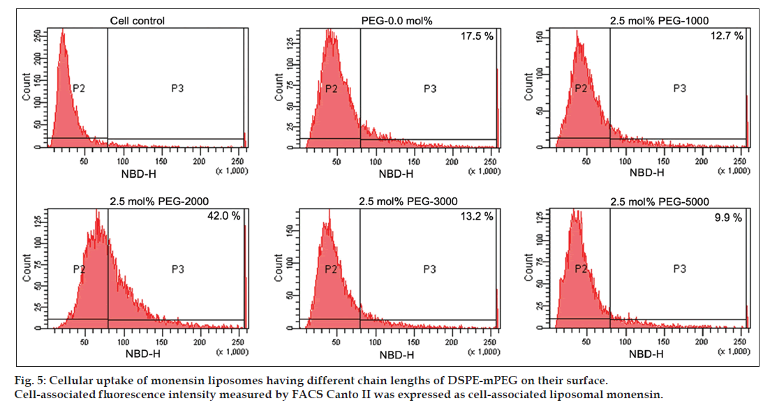
Cellular uptake of neutral conventional and various sterically stabilized liposomal formulations of monensin was assessed using NBD‑PC (1‑palmitoyl ‑2‑{12‑[(7‑nitro‑2‑1,3‑benzoxadiazol‑4‑yl)amino] lauroyl}‑sn‑glycero‑3‑phosphocholine) as fluorescent surface marker. Briefly, cells at a density of 8×10⁵ cells/well/1 ml were plated in 24 well plates, 24 h before the experiment. The monolayer cultures were washed twice with DBSS and incubated with or without 50 nM monensin intercalated in liposomes containing 0‑7.5 mol% DSPE‑mPEG of different chain length (1000, 2000, 3000 and 5000) at a final lipid concentration of 20‑25 nM/ml for 1 h at 37°. Then, varying concentrations of liposomal ricin in 100 µl DBSS was added and cells were incubated at 37° for 4 h. Cells were then washed thrice with ice cold DBSS to remove unbound liposomes. The cells were fixed with 2% paraformaldehyde, detached by a brief trypsin‑EDTA (ethylenediaminetetraacetic acid) treatment and the fluorescence intensity in cells was analysed by fluorescence activated cell sorter (FACS Canto II, BD Bioscience, USA). Cellular uptake of the liposomal formulations was quantified on the basis of fluorescence intensity associated with the cells.
Results and Discussion
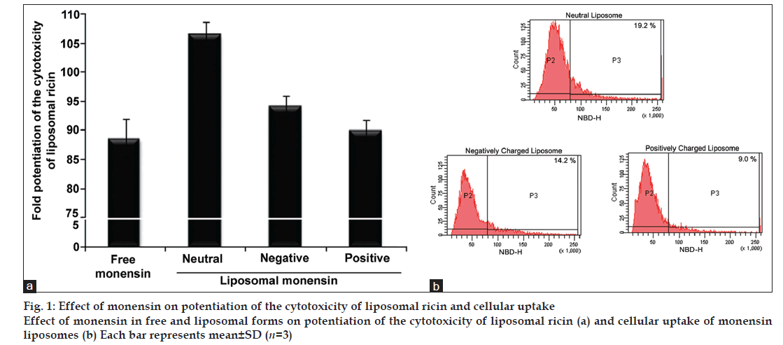
In the present investigation, monensin was intercalated in neutral, negatively and positively charged liposomes and its ability to potentiate the cytotoxicity of ricin entrapped in negatively charged sterically stabilised liposomal form was evaluated in KB cells. These results of liposomal monensin were compared with free monensin to develop an optimal delivery system for liposomal ricin in combination with monensin for selective elimination of tumour cells. The extent of enhancement of cytotoxicity of ricin by monensin intercalated in various charged liposomes is found to be dependent on the lipid composition of monensin liposomes and significantly higher as compared to free monensin. Monensin exhibit maximum enhancing ability (106.5 fold) when delivered through intercalated into neutral liposomes followed by negatively (94.2 fold) and positively charged (90 fold) liposomes as compared to free monensin (88.6 fold) (fig. 1a). The cellular uptake studies of monensin liposomes clearly indicate that there is a direct correlation between the enhancements of the cytotoxicity of ricin encapsulated into sterically stabilised liposomes by monensin intercalated into various charged liposomes with their amount of cell association (fig. 1b). These results are highly comparable with earlier reports that interaction of liposomes with mammalian cells is highly dependent on several factors like lipid composition, size, charge, bilayer fluidity, hydrophilicity of the liposomes as well as cell types [18,19]. As our results clearly showed that liposomes can be used as delivery vehicle for monensin to enhance the cytotoxicity of liposomal ricin and as neutral monensin liposome has the maximum ability to enhance the cytotoxicity of liposomal ricin, all subsequent work was therefore carried out using monensin intercalated into neutral liposomal formulations.
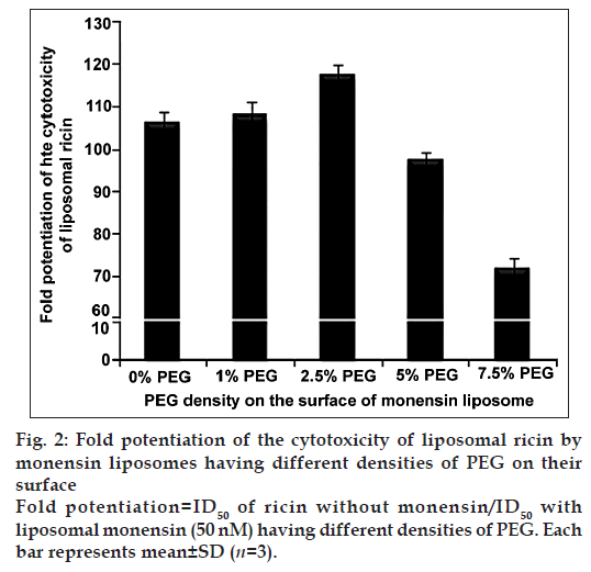
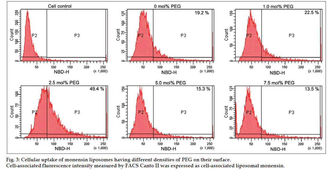
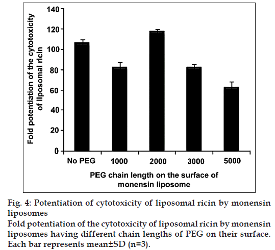
Studies on the effect of various densities of DSPE‑mPEG‑2000 on the surface of neutral monensin liposomes revealed that monensin exhibits maximum enhancement (117.6 fold) of liposomal ricin cytotoxicity at 2.5 mol% DSPE‑mPEG‑2000 on the surface (fig. 2). In order to evaluate whether the differential efficacy exhibited by liposomal monensin on the enhancement of the cytotoxicity of liposomal ricin is due to the difference in cellular uptake, the cellular uptake of monensin liposomes at 37° in KB cells was examined. Fig. 3 shows the cellular uptake of monensin liposomes having different densities of DSPE‑mPEG‑2000 as expressed in the form of percentage of fluorescence of KB cells studied by FACS. Cellular uptake studies and measurement of zeta potential of monensin liposomes having various densities of DSPE‑mPEG‑2000 on the surface revealed that there is high correlation between the enhancement of the cytotoxicity of ricin with their amount of cell association and zeta potential on the surface of monensin liposomes. The maximum enhancement of the cytotoxicity of liposomal ricin was observed when the cellular uptake of liposome was maximum (49.4%) and the zeta potential value on the surface of liposomes was −0.645 at 2.5 mol% DSPE‑mPEG‑2000 on the surface of liposomes. Keeping 2.5 mol% DSPE‑mPEG as constant, we varied the chain length of PEG on the surface of monensin liposomes and examined the efficacy of monensin in these liposomes on the enhancement on the cytotoxicity of liposomal ricin in KB cells (fig. 4). It was observed that the PEG chain length of 2000 has maximum influence on the ability of the monensin in neutral PEG‑liposomes to enhance the cytotoxicity of liposome entrapped ricin. The liposomal ricin cytotoxicity enhancing potency of monensin in PEG‑liposomes is reduced from 117.6 fold to 82.4 and 63.0 fold when chain length of PEG was increased from 2000 to 3000 and 5000. It was also observed that the fold enhancement of the cytotoxicity of liposomal ricin was again reduced (from 117.6 to 82.4 fold) on reducing the length of PEG from 2000 to 1000. The maximum enhancement of the cytotoxicity of liposomal ricin by PEG‑liposomal monensin was observed when the cellular uptake of liposome was maximum (42.0%) and the zeta potential value on the surface of liposomes was −0.645 at 2.5 mol% DSPE‑mPEG of chain length 2000 on the surface of liposomes (fig. 5). These results clearly showed that the value of zeta potential on the surface of PEG liposomes is crucial for expression of the biological activity of monensin in PEG‑liposomes and the optimum value is around −0.645. These results are in agreement with earlier reports that the interaction of liposomes with cancer cells not only depends on the surface charge, but also on density, chain length and lipid anchor of PEG‑lipid [14]. This zeta potential on the surface of liposomes can be modulated by two ways, either by changing the density of DSPE‑mPEG‑2000 or by changing the chain length of PEG in DSPE‑mPEG on the surface of liposomes. This report is the first of its kind to show that liposomal monensin is very effective in enhancing the cytotoxicity of liposomal ricin in human cancer cells and liposome can be used as in vivo delivery vehicle to eliminate tumour cells.
References
- Endo Y, Mitsui K, Motizuki M, Tsurugi K. The mechanism of action of ricin and related toxic lectins on eukaryotic ribosomes. The site and the characteristics of the modification in 28 S ribosomal RNA caused by the toxins. J Biol Chem 1987;262:5908-12.
- Tyagi N, Rathore SS, Ghosh PC. Enhanced killing of human epidermoid carcinoma (KB) cells by treatment with ricin encapsulated into sterically stabilized liposomes in combination with monensin. Drug Deliv 2011;18:394-404.
- Rathore SS, Ghosh PC. Effect of surface charge and density of distearylphosphatidylethanolamine-mPEG-2000 (DSPE-mPEG-2000) on the cytotoxicity of liposome-entrapped ricin: Effect of lysosomotropic agents. Int J Pharm 2008;350:79-94.
- Bharadwaj S, Rathore SS, Ghosh PC. Enhancement of the cytotoxicity of liposomal ricin by the carboxylic ionophore monensin and the lysosomotropic amine NH4Cl in Chinese hamster ovary cells. Int J Toxicol 2006;25:349-59.
- Vasandani VM, Madan S, Ghosh PC. In vivo potentiation of ricin toxicity by monensin delivered through liposomes. Biochim Biophys Acta 1992;1116:315-23.
- Madan S, Ghosh PC. Enhancing potency of liposomal monensin on ricin cytotoxicity in mouse macrophage tumor cells. Biochem Int 1992;28:287-95.
- Madan S, Vasandani VM, Ghosh PC. Potentiation of ricin cytotoxicity by liposomal monensin under in vitro and in vivo conditions. Indian J Biochem Biophys 1993;30:405-10.
- Griffin T, Rybak ME, Recht L, Singh M, Salimi A, Raso V. Potentiation of antitumor immunotoxins by liposomal monensin. J Natl Cancer Inst 1993;85:292-8.
- Shaik MS, Jackson TL, Singh M. Effect of monensin liposomes on the cytotoxicity of anti-My9-bR immunotoxin. J Pharm Pharmacol 2003;55:819-25.
- Singh M, Griffin T, Salimi A, Micetich RG, Atwal H. Potentiation of ricin A immunotoxin by monoclonal antibody targeted monensin containing small unilamellar vesicles. Cancer Lett 1994;84:15-21.
- Zalipsky S, Brandeis E, Newman MS, Woodle MC. Long circulating, cationic liposomes containing amino-PEG-phosphatidylethanolamine. FEBS Lett 1994;353:71-4.
- Allen TM, Austin GA, Chonn A, Lin L, Lee KC. Uptake of liposomes by cultured mouse bone marrow macrophages: Influence of liposome composition and size. Biochim Biophys Acta 1991;1061:56-64.
- Levchenko TS, Rammohan R, Lukyanov AN, Whiteman KR, Torchilin VP. Liposome clearance in mice: The effect of a separate and combined presence of surface charge and polymer coating. Int J Pharm 2002;240:95-102.
- Sadzuka Y, Kishi K, Hirota S, Sonobe T. Effect of polyethyleneglycol (PEG) chain on cell uptake of PEG-modified liposomes. J Liposome Res 2003;13:157-72.
- Borowik T, Widerak K, Ugorski M, Langner M. Combined effect of surface electrostatic charge and poly (ethyl glycol) on the association of liposomes with colon carcinoma cells. J Liposome Res 2005;15:199-213.
- Madan S, Ghosh PC. Monensin intercalation in liposomes: Effect on cytotoxicities of ricin, Pseudomonas exotoxin A and diphtheria toxin in CHO cells. Biochim Biophys Acta 1992;1110:37-44.
- Stewart JC. Colorimetric determination of phospholipids with ammonium ferrothiocyanate. Anal Biochem 1980;104:10-4.
- Gregoriadis G. Engineering liposomes for drug delivery: Progress and problems. Trends Biotechnol 1995;13:527-37.
- Ryman BE, Tyrrell DA. Liposomes – Bags of potential. Essays Biochem 1980;16:49-98.
