- *Corresponding Author:
- Sultan Areshi
Department of Biology, College of Science, Jazan University, Jazan 45142, Kingdom of Saudi Arabia
E-mail: smareshi3@gmail.com
| Date of Received | 02 Jun 2025 |
| Date of Revision | 03 Jun 2025 |
| Date of Accepted | 15 July 2025 |
| Indian J Pharm Sci 2025;87(4):126-132 |
This is an open access article distributed under the terms of the Creative Commons Attribution-NonCommercial-ShareAlike 3.0 License, which allows others to remix, tweak, and build upon the work non-commercially, as long as the author is credited and the new creations are licensed under the identical terms
Abstract
Shrimp are highly susceptible to diseases caused by microorganisms, and this greatly affects productivity. Recently, studies investigating shrimp diseases have increased, and this may be due to the high density and the intensive greenhouse model. This study aims to isolate and diagnose fungi found in prawn shrimp (Penaeus monodon) in Jizan Sea. We examined and isolated fungi from prawn shrimp (Penaeus monodon) into different locations in the body (head, abdomen, and tail). We found one type of fungi (Fusarium solani) in most body sites, especially in the abdomen and head area observed under the microscope. This type of fungi is highly invading the whole body of prawn shrimp, which may cause deterioration in the lives of shrimp in the near future and transmit many diseases to its users.
Keywords
Binary matrix, accessions, phenogram, parasite, fungi
It has been found that shrimp are highly susceptible to diseases such as fungal diseases caused by microorganisms, and this may affect productivity[1]. Few studies have revealed some of the factors affecting invertebrates, and only a few viruses, bacteria and protozoa are known to cause disease in these crustaceans (e.g. shrimp)[2]. Recently, studies investigating shrimp diseases have increased, and this may be due to the high density and intensive greenhouse model[3,4], and this may be a cause of significant economic losses every year[5]. Therefore, it is important that we quickly develop a strategy for diagnosing shrimp diseases. The presence of fungi in the body of shrimp may lead to negative effects on their health, growth, and behaviour, including yeast (Candida spp.): Candida spp. It is a common fungus that infects shrimp. It causes infections in the mucous membranes and skin and may affect the health and growth of shrimp. Fusarium spp. (Sordariomycetes class, Hypocreales order, Nectriaceae family) is a widely distributed in plants, soil, freshwater, and brackish water[6,7]. This fungus causes infections in the skin and cortical parts of shrimp and affects the functions of internal organs. Lagenidium spp. Parasitic to shrimp. They infect skin tissue and cause serious infections that affect the health of shrimp[1,2]. A study focused on the accumulative mortalities of white shrimp Penaeus vannamei (P. vannamei) of black spot disease by Fusarium solani (F. solani), which showed that the histopathological characterization of the relationship between P. vannamei against F. solani are hemolytic infiltration, encapsulation, melanization, etc. This can cause melting failure and contribute to the death of the sick individuals[8]. The topographical investigation of black melanin pigment of white shrimp infected by F. solani resulted in altered immune response and histopathological effect[9].
This study aims to isolate and diagnose fungi found in prawn shrimp (Penaeus monodon) (P. monodon)) in Jizan Sea. We examined and isolated fungi from prawn shrimp (P. monodon) into different locations in the body (head, neck, abdomen, and tail). In addition, we cultivated the fungi we obtained in different dishes and precisely identified their types and the extent of their harm to shrimp to develop effective control strategies the spread of these organisms and reducing their negative impact on the health and sustainability of shrimp farming. This research will also contribute to increasing awareness of the importance of fungi and parasites research in the field of aquaculture and marine resource conservation.
Materials and Methods
Prawn collection and identification:
100 samples of prawn shrimp (P. monodon) were collected from Jazan Sea from different random spots near and far the seashore on the middle of September. All samples kept into ice bag after collection and return to the laboratory to examine morphological characters and fungi parasites. We measured the length (mm) of each prawn shrimp sample using the foot and the antennae (mm) as well. Then we measured the weight (grams) using an electronic scale for each sample. Next, we extracted tissue from the head, abdomen and tail of five shrimp samples and placed them in a petri dish containing agar separately (each petri dishes has one extracted tissue from one species). Plates were placed in the incubator for a period of 8 d.
Fungi isolation:
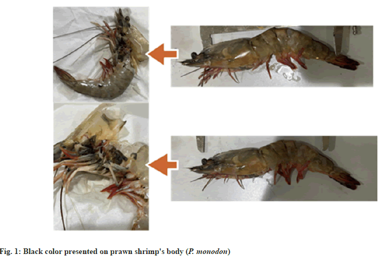
On the 9th d, we extracted samples from the areas where the fungi had grown using small tweezers and placed them on sterile microscopic slides with the addition of Safranine O dye. We put a mark on each slide to know which part of the body contains the three samples, whether the head, abdomen, or tail. Then we identified the types of fungi extracted under the microscope (fig. 1).
Scoring data:
Morphological characters were determined for 15 accessions. Some characters were qualitative and quantitative. All data were scored as a binary matrix in the form of (0; absent, 1; present). The phenogram was established to differentiate among different characters of 15 accessions. Similarity matrix was performed to evaluate the degree of relations among accessions[10].
Results and Discussion
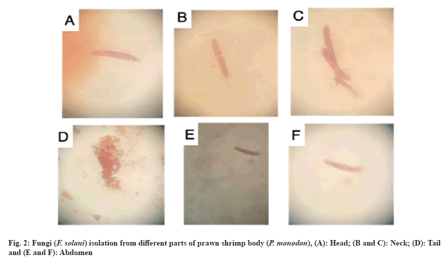
We found one type of fungi (F. solani) is extremely invaded the shrimp body. This fungi was found into different body location, but we mostly found high number of this fungi in the head and abdomen under the microscope. This was observed in the microscope (F. solani). The fungi was widely spread in the body, especially where we saw black color in the body (see fig. 1), which contained these fungi, in compare to those shrimp that were completely white, but most of the shrimp showed fungi in the body. In the tail area, we found the same type of fungi, but at the first growth stage, unlike the head and abdomen area, where the fungal growth was completely grown (fig. 2).
Morphological characters were qualitative as body color and specific character, dark head, dark tail and dark abdomen. Quantitative characters were regarded as measured ones as frequency (%), average body length (mm), average antenna length (mm) and average body weight (g) (Table 1).
| Accession No. | Frequency (%) | Average body length (mm) | Average antenna length (mm) | Average body weight (g) | Body color | Specific character | Parasite |
|---|---|---|---|---|---|---|---|
| 1 | 5 | 10.28 | 7.64 | 33.56 | Light prawn | NA | Absent |
| 2 | 3 | 8.5 | 0 | 29.36 | Light prawn | No antenna | Absent |
| 3 | 1 | 7.5 | 8.4 | 38.25 | Light prawn | Dark tail | Present |
| 4 | 2 | 7.55 | 6.25 | 27.36 | Light prawn | Dark tail | Absent |
| 5 | 37 | 8.72 | 9.07 | 28.41 | Dark prawn | NA | Absent |
| 6 | 11 | 8.4 | 5.3 | 27.05 | Dark prawn | Dark abdomen | Absent |
| 7 | 1 | 10.9 | 5.4 | 32.44 | Light prawn | Dark head | Absent |
| 8 | 3 | 7.7 | 0 | 34.53 | Dark prawn | No antenna | Absent |
| 9 | 1 | 6 | 8.8 | 25.6 | Light prawn | NA | Present |
| 10 | 12 | 7.76 | 9 | 29.82 | Light prawn | NA | Absent |
| 11 | 8 | 8.5 | 8.69 | 25.17 | Dark prawn | Dark head | Absent |
| 12 | 7 | 10.4 | 9.5 | 23.21 | Dark prawn | Dark tail | Absent |
| 13 | 1 | 6.5 | 5.3 | 30.42 | Light prawn | Dark abdomen | Absent |
| 14 | 1 | 6 | 11.6 | 33.9 | Dark prawn | Dark head | Present |
| 15 | 7 | 6.9 | 8.36 | 37.38 | Dark prawn | NA | Present |
Table 1: Morphological Characters of Prawn Shrimp Accessions in the Sample
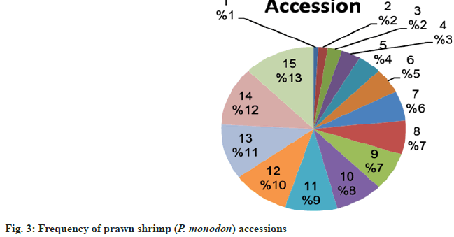
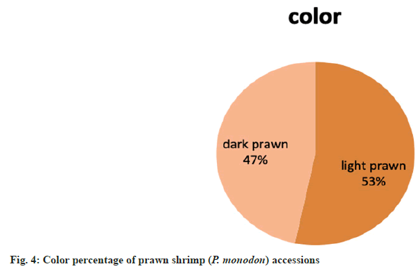
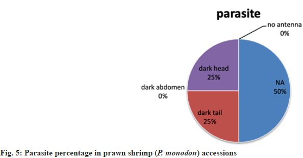
Accession no. 5 was recorded as the highest frequency in the sample while accession no. 3, 7, 9, 13 and 14 were recorded as the lowest one (fig. 3). Most predominant body color was light (53 %) fig. 4. Fungi parasite was not present in all accessions. Accessions, whose fungi parasites occupied their body and had not any specific character, scored the half frequency of the sample. Accessions with specific characters like no antenna and dark abdomen had not.
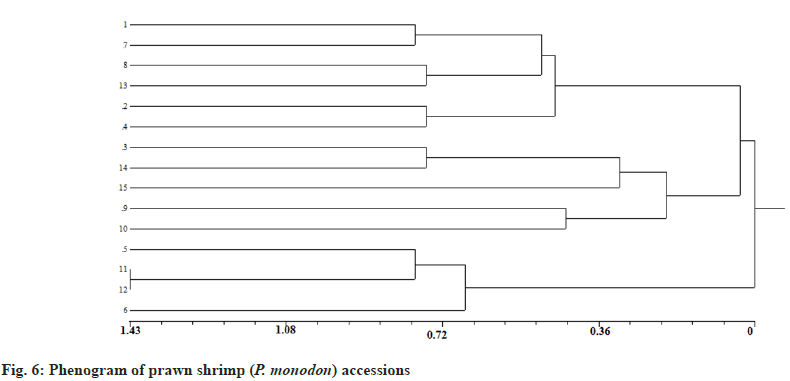
Binary matrix in presence of 0 and 1 parameters established the phenogram on similarities and dissimilarities among accession morphology (Table 2). The phenogram represented all accessions in three main groups; the first group included accession no. 1, 2, 4, 7, 8 and 13 while second group included 3, 9, 10, 14 and 15 finally, the third one delimited only with 5, 6, 11 and 12 (fig. 5). A similar matrix indicated that accession no. 11 and 12 were scored highest value while accession no. 2 and 15 were scored lowest value (Table 3 and fig. 6).
| Accession No. | Frequency | Average body length | Average antenna length | Average body weight (g) | Body color | Specific character | Parasite |
|---|---|---|---|---|---|---|---|
| 1 | 0 | 1 | 0 | 1 | 1 | 0 | 0 |
| 2 | 0 | 1 | 0 | 0 | 1 | 1 | 0 |
| 3 | 0 | 0 | 1 | 1 | 1 | 1 | 1 |
| 4 | 0 | 0 | 0 | 0 | 1 | 1 | 0 |
| 5 | 1 | 1 | 1 | 0 | 0 | 0 | 0 |
| 6 | 1 | 1 | 0 | 0 | 0 | 1 | 0 |
| 7 | 0 | 1 | 0 | 1 | 1 | 1 | 0 |
| 8 | 0 | 0 | 0 | 1 | 0 | 1 | 0 |
| 9 | 0 | 0 | 1 | 0 | 1 | 0 | 1 |
| 10 | 1 | 0 | 1 | 0 | 1 | 0 | 0 |
| 11 | 1 | 1 | 1 | 0 | 0 | 1 | 0 |
| 12 | 1 | 1 | 1 | 0 | 0 | 1 | 0 |
| 13 | 0 | 0 | 0 | 1 | 1 | 1 | 0 |
| 14 | 0 | 0 | 1 | 1 | 0 | 1 | 1 |
| 15 | 1 | 0 | 1 | 1 | 0 | 0 | 1 |
Table 2: Binary Matrix of Prawn Shrimp (p. Monodon) Accessions
| Sr. no. | 1 | 2 | 3 | 4 | 5 | 6 | 7 | 8 | 9 | 10 | 11 | 12 | 13 | 14 | 15 |
|---|---|---|---|---|---|---|---|---|---|---|---|---|---|---|---|
| 1 | |||||||||||||||
| 2 | e | ||||||||||||||
| 3 | c | c | |||||||||||||
| 4 | d | f | d | ||||||||||||
| 5 | c | c | a | b | |||||||||||
| 6 | c | e | a | d | e | ||||||||||
| 7 | f | f | d | e | b | d | |||||||||
| 8 | d | d | d | e | b | d | e | ||||||||
| 9 | c | c | e | d | c | a | b | b | |||||||
| 10 | c | c | c | d | e | c | b | b | e | ||||||
| 11 | b | d | b | c | f | f | c | c | b | d | |||||
| 12 | b | d | b | c | f | f | c | c | b | d | 1 | ||||
| 13 | e | e | e | f | a | c | f | f | c | c | b | b | |||
| 14 | b | b | f | c | b | b | c | e | d | b | b | c | b | ||
| 15 | b | 0 | d | a | d | b | a | c | d | d | c | c | b | e |
Table 3: Similarity Matrix of Prawn Shrimp (p. Monodon) Accessions, (a); 0.14; (b): 0.28; (c): 0.43; (d): 0.57; (e): 0.71 and (f): 0.86
Many types of fungi can be present in the body of shrimp, such as dermatophytes and enteritis fungi. These fungi are usually found in the aquatic environment in which shrimp live and may infect the shrimp?s body when their immunity is weak or tired. The effect of these fungi on shrimp can be diverse, as they can cause infections of the skin and intestines and affect the functions of their vital organs, which may lead to weakness. Growth and death in severe cases. Investing a new fungi (F. solani) prawn shrimp (P. monodon) may lead to further threaten of shrimp's life.
The presence of fungi in different location of the body can explain that this fungi is spreading out fast as we noticed black color around the whole body of most shrimps. This negatively affect its health, growth and behaviour. Liang et al.[8] reported that F. solani was regarded as haemocytic agent which contributed the molting failure and spread the prawn mortality. These organisms can cause infections in the mucous membranes and shell parts of shrimp, deteriorating their health and affecting their behaviour. After that it can penetrate later from prawn to consumer?s central nervous system through meningitis and cerebrospinal fluid caused epidural anaesthesia[11].
This may affect his ability to feed and breathe properly, and therefore may affect his growth and ability to survive. In short, shrimp are a small, highly nutritious marine organism that is very popular in the food and seafood industry. However, there are different types of fungi and parasites that can infect shrimp and affect their health and growth. The effects of these organisms may range from skin and intestinal infections to effects on his vital systems.
From the current study, accessions no. 5 and 10 exhibited the highest frequency that referring to be more dominant and familiar to Jazan nature. Highest percentage of parasitism in accessions lacking specific characters indicated that these characters provided the accessions more immunity against parasitism. Accessions no. 11 and 12 expressed the highest related even though they were not the dominant ones reflecting that they may be invaded out the side the area not belonging to the original ancestors. The distantly relation between accession no. 2 and 15 emphasized the previous investigations. This study stated that genetic variation approaches should complete the work to give the complete pictures about endemic and invaded accessions.
By studying the impact of fungi on shrimp, we can develop strategies to reduce the spread of these organisms and protect the health and sustainability of shrimp farming. This study will also increase awareness of the importance of studying fungi and parasites in the field of aquaculture and marine resource conservation. To maintain the health of shrimp, it is recommended to pay attention to the cleanliness of the water and environment in which the shrimp live and to take appropriate preventive measures to limit the transmission of fungi and parasites to it.
The current study of the prawn accessions reflected the degree of human impact on marine environment. The phylogenetic analysis among prawn accessions was the indicator of origin accession and developmental changes with the same species throughout coastal countries and different seas. This investigation confirmed on the danger of F. solani not only on marine creatures but may involve in other living organisms like humans.
Acknowledgments:
Author acknowledge Jazan University and Dr. Said Alkateib for providing research facilities.
Author's contribution:
The experiment such laboratory work, data collection, statistical analyses, and manuscript written were conducted by Said Alkateib at Biology Department of Jazan University.
Conflict of interests:
The authors declared no conflict of interests.
References
- Silva LR, Souza OC, Fernandes MJ, Lima DM, Coelho RR, Souza-Motta CM. Culturable fungal diversity of shrimp Litopenaeus vannamei boone from breeding farms in Brazil. Braz J Microbiol 2011;42(1):49-56.
- Pontes CS, Arruda MD. Behaviour of Litopenaeus vannamei (Boone) (Crustacea, Decapoda, Penaeidae) in relation to artificial food offer along light and dark phases in a 24 h period. Rev Bras Zoo 2005;22(3):648-52.
- Leung AA, McAlister FA, Finlayson SR, Bates DW. Preoperative hypernatremia predicts increased perioperative morbidity and mortality. Am J Med 2013;126(10):877-86.
[Crossref] [Google Scholar] [PubMed]
- Akonor PT, Ofori H, Dziedzoave NT, Kortei NK. Drying characteristics and physical and nutritional properties of shrimp meat as affected by different traditional drying techniques. Int J Food Sci 2016;2016(1):7879097.
[Crossref] [Google Scholar] [PubMed]
- Chaijarasphong T, Munkongwongsiri N, Stentiford GD, Aldama-Cano DJ, Thansa K, Flegel TW, et al. The shrimp microsporidian Enterocytozoon hepatopenaei (EHP): Biology, pathology, diagnostics and control. J Invertebr Pathol 2021;186:107458.
[Crossref] [Google Scholar] [PubMed]
- Le VK, Hatai K, Yuasa A, Sawada K. Morphology and molecular phylogeny of Fusarium solani isolated from kuruma prawn Penaeus japonicus with black gills. Fish Pathol 2005;40(3):103-9.
- Palmero D, Iglesias C, De Cara M, Lomas T, Santos M, Tello JC. Species of Fusarium isolated from river and sea water of southeastern Spain and pathogenicity on four plant species. Plant Dis 2009;93(4):377-85.
[Crossref] [Google Scholar] [PubMed]
- Yao L, Wang C, Li G, Xie G, Jia Y, Wang W, et al. Identification of Fusarium solani as a causal agent of black spot disease (BSD) of Pacific white shrimp, Penaeus vannamei. Aquaculture 2022;548:737602.
- Mansour AT, Eldessouki EA, Khalil RH, Diab AM, Selema TA, Younis NA, et al. In vitro and in vivo antifungal and immune stimulant activities of oregano and orange peel essential oils on Fusarium solani infection in whiteleg shrimp. Aquaculture Int 2023;31(4):1959-77.
- Atta E, El-Shabasy A. Chemotaxonomic and morphological classification of six Indigofera species in Jazan region, KSA. J Saudi Chem Soc 2022;26(3):101476.
- Baum SG. Fusarium solani meningitis related to medical tourism—a clinical update. NEJM J Watch 2024:NA57091.
