- *Corresponding Author:
- P. Ghosh
Natural Product and Polymer Chemistry Laboratory, Department of Chemistry, University of North Bengal, Darjeeling – 734 013
E-mail: pizy12@yahoo.com
| Date of Submission | 27 August 2009 |
| Date of Decision | 8 March 2010 |
| Date of Acceptance | 4 August 2010 |
| Indian J Pharm Sci, 2010, 72 (4): 504-507 |
Abstract
In continuation of our studies on the phytochemical investigation of medicinal plants available in the foothills of Darjeeling and Teri, we report herein the isolation of two triterpenoids betulinic acid and lupeol from the leaf extract of Psidium guajava and their potential antimicrobial and phytotoxic activities. All the structures of the isolated compounds were confi rmed by spectral (IR, NMR) analysis and by comparison with the literature reports.
Keywords
Betulinic acid, lupeol, minimum inhibitory concentration, myrtaceae, Psidium guajava, triterpenoid
The Himalayan region of Darjeeling and Terai are rich in bio diversity with plants having pronounced medicinal activities as evidenced by recent literature reports [1-3] as well as by the tribal medicinal practice in this region. Plants of the family Myrtaceae are extensively used in indigenous medicine from prehistoric ages. Psidium guajava is an important representative of this family. Present day reports about P. guajava are attracting because of their highly encouraging biological activities [3-11]. Different parts of these plants are used in the traditional system of medicine for the treatment of various human ailments such as ulcers, bronchitis, eye sores, bowels, diarrhoes and cholera [3-6]. It is reported in the literature that the leaf extract of P. guajava has antitussive, antibacterial, hemostatic, antioxidant and narcotic properties [7-10]. Recently Abreu et al, have reported that guava extract can alter the labelling of blood with technetium- 99m [11].
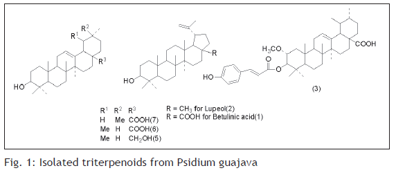
In view of the attributed medicinal properties and in an ongoing search for bioactive triterpenoids from plants of Myrtaceae available in Darjeeling foothills, the toluene extract of leaves of P. guajava was selected for further investigation. The leaf extract of P. guajava was found to contain two new triterpenoids (1 and 2) along with earlier reported guajanoic acid (3) [6], β-sitosterol (4), uvaol (5), ursolic acid (6) and oleanolic acid (7)(fig. 1). Compounds 1 and 2 have been characterized as betulinic acid and lupeol respectively. This is the first report of the isolation of these two triterpenoids from the leaf extract of P. guajava available plenty in the foothills of Darjeeling. In addition to that preliminary studies towards the antimicrobial and phytotoxic activities of these two compounds, which have not yet reported so far from this source, have also been carried out against some fungal and bacterial pathogens.
All the melting points were determined by open capillary method and are uncorrected. The NMR spectra were recorded in CDCl3 solutions at ambient temperature on a Bruker Avance 300 MHz-FT NMR spectrometer using 5 mm BBO probe. The chemical shift δ are given in ppm related to tetra methyl silane (TMS) as internal standard. The coupling constant (J) are reported in Hz. The IR spectra were recorded in Shimadzu FT-IR spectrophotometer in KBr discs.
Fresh leaves of P. guajava were collected in bulk from young mature plants at the Sukna belt of Darjeeling foothills during early summer, washed, shade dried and milled into coarse powder by a mechanical grinder. The prepared powdered leaves were then used for further studies. The powdered plant material was extracted with toluene using Soxhlet apparatus for 72 h. The solvents were then removed under reduced pressure and a sticky brown residue was obtained. This residue was then purified by column chromatography using silica gel (60-120) mesh and suitable proportions of petroleum ether and ethyl acetate were used as the eluent.
In this present work the in vitro antifungal antibacterial activities and the phytotoxicity of the two isolated triterpenoids have been studied. Five different fungal pathogens namely, Calletotricheme camellie, Fussarium equisitae, Alterneria alternate, Curvularia eragrostidies, Colletrichum Gleosproides were used for the antifungal study. For antibacterial study Escherichia Coli, Bacillus Subtilis, Staphylococcus aureus, Enterobactor were used as bacterial pathogen. Suitable strains of these organisms were procured from the microbiology laboratory of our institute. MICs (minimum inhibitory concentration) of the triterpenoids against bacterial and fungal pathogens are reported in Tables 1 and 2, respectively. DMSO (dimethyl sulfoxide) was used as solvent to prepare different concentrations of the triterpenoids. Solvent control (DMSO) was also maintained throughout the experiment. All experiments were performed in petridishes and were incubated at 37o for 48 h.
| Compounds | MIC in μg/ml against different strains of bacteria | ||||
|---|---|---|---|---|---|
| EC | BS | SA | EB | ||
| 1 | 150 | <100 | 100 | 100 | |
| 2 | 200 | 100 | 200 | 100 | |
| Ampicillin | 128 | 64 | 64 | 128 | |
BS- Bacillus. subtilis, EC- Escherichia coli, SA- Staphylococcus aureus, EB-Enterobactor, MIC- Minimum inhibitory concentration
Table 1: Mic of 1 and 2 against Different Bacteria
| Compounds | MIC in μg/ml against different fungi | ||||
|---|---|---|---|---|---|
| CG | FE | CE | AA | CC | |
| 1 | <5 | 2.5 | 10 | 5 | <5 |
| 2 | 10 | 5 | 10 | 5 | 5 |
| Streptomycin | 1.25 | 2.5 | <2.5 | 2.5 | 2.5 |
CG- Colletrichum gleosproides, FE- Fussarium equisitae, CE- Curvularia eragrostidies, AA- Alterneria alternate, CC- Calletotricheme camellie.
Table 2: Mic of 1 and 2 against Different Fungi
The bacterial growth was confirmed by a change of yellow to purple colour. Bacterial nutrient media was prepared by using agar, beef extract and bacto peptone in distilled water and the pH of the solution (6.8-7.0) was adjusted. Culture media for fungal strains were prepared by mixing in suitable proportions of potato extract, dextrose and agar powder. All glass apparatus, culture media were autoclaved before use. The whole process was carried out in inoculation chamber. Additionally slide germination method was also used for determination of antifungal activity [12] (Table 3). The antifungal activities between these compounds and streptomycin and antibacterial activity with ampicillin, a β-lactam antibiotic were compared.
| FungalPathogen | Betulinic acid | Lupeol | ||||
|---|---|---|---|---|---|---|
| PGa | PI | ALb(μm) | PGa | PI ALb(μm) | ||
| CC | 00 | 100 | 00 | 0.5 | 95 | 4.5 |
| FE | 00 | 100 | 00 | 00 | 100 | 00 |
| AA | 00 | 100 | 00 | 00 | 100 | 00 |
| CG | 00 | 100 | 00 | 00 | 100 | 00 |
| CE | 00 | 95 | 6.0 | 10 | 95 | 9.0 |
CG- Colletrichum Gleosproides, FE- Fussarium equisitae, CE- Curvularia eragrostidies, AA- Alterneria alternate, CC- Calletotricheme camellie. PG-Percent germination, PI-Percent Inhibition, AL-Average germ tube length, aBased on 200 spores, bBased on 25 germ tubes. All data were taken after 48 h of incubation.
Table 3: Antifungal Properties of 1 and 2 Based on Spore Germination Bioassay
For studying the inhibitory effect [12] of the two triterpenoids against test fungal pathogens following slide germination method, the spores of the pathogens were allowed to germinate in presence of the prepared and the 50% ethanol extracts. Compound solution was placed on the centre of the grease free microscope slide. In control the corresponding solvent, either sterile distilled water or 50% ethanol was placed. Thirty microlitre spore suspension prepared from ten days culture of the fungal pathogens were added to the spots in both experimental and control slides. In case of 50% ethanol extract, spore suspension was added after ethanol was evaporated. Three experimental slides were taken for each compound. The slides were then incubated at 28o in a humid chamber. Two small glass rods (60 mm in length) were placed in a 90 mm Petri dish and a slide was placed on the rods in a uniformly balanced position. Then the Petri dish was filled with sterile distilled water so that the bottom of the slide remained just above the water surface. The petridish was then covered and incubated at 28o. Following 48 h of incubation, the slides were stained with lacto phenol cotton blue mixture and observed in each slide for germination. Numbers of aspersoria formed were also observed and lengths of 50 germ tubes were measured. The entire experiment was repeated thrice.
Seeds of rice (Oriza sativa), wheat (Triticum aestirium), and pea (Pisum sativum) were collected from local market. The assay seeds were shorted for uniformity of size and all damaged seeds were discarded. Before the bioassay seeds were washed with tap water and the surface were sterilized using NaCl (10% v/v) for 10 min followed by several washes in sterile distilled water. For testing phytotoxicity dehydrated ethanol was used as control. Bioassays were carried out using petridishes (90 mm diameter) containing a sheet of Whatman 1 filter paper as support. Test solutions (5 ml) was added to the filter paper in the petridish and dried completely in vacuo at 40o. Five seeds from each category were placed on the filter paper and incubated for 7 days at 25o in the dark. The effects of the pure compounds were determined by measuring the elongation of roots and averaged for each concentration.
Compound (1) was isolated as white crystals (CHCl3+MeOH) of m.p. 299-301o. IR spectrum has exhibited hydroxyl at vmax 3610, 1020 cm-1 and exomythylene at vmax 3060, 1630, 880 cm-1. The 1H NMR spectrum revealed signals for five tertiary methyls δH 0.65, 0.75, 0.90, 0.96 and 0.98, a vinyl methyl δH=1.97 broad d, J= 0.5 Hz), a secondary carbinol δH=3.16 dd, J= 9.5 and 6.0 Hz) and δH= 2.95 (ddd, J= 9.0, 6.0 and 0.5 Hz) an exomethylene group δH=4.55 (1H, d, J= 0.4 Hz) and δH= 4.65(1H, d, J= 0.4 Hz). These data indicated a pentacyclic triterpenoid of betulinic acid, confirmed by comparison with already published data [13-16]. The 13C NMR spectrum of (1) showed six methyl group at δc 27.9 (C-23), 15.4 (C-24), 16.2 (C-25), 16.3 (C-26), 14.6 (C-27), 19.6 (C-30) and exomethylene group at δc 150.0 (C-30), 108.8 (C-29) and a secondary carbon bearing hydroxyl at δc 79.0 (C-3) and a carboxyl group at δc= 180.6 (C-28) in addition to ten methylene, five methine and five quaternary carbons. These data were identical to those reported for betulinic acid [13-16].
Lupeol (2) was isolated as white crystals from CHCl3+MeOH mixture and gave m.p. 210-21 D = +30.4 (conc. 0.58 in CHCl3). Its IR spectrum exhibited hydroxyl at vmax 3610, 1020 cm-1 and exomethylene at vmax 3070, 1640, 887 cm-1 absorption. The 1H NMR exhibited six tertiary methyl signals at H 0.75, 0.77, 0.80, 0.92, 0.94 and 1.02, a vinyl methyl group at vmax 1.66 (broad d J= 0.5 Hz)], a secondary carbinol group at 3.20 (dd, J= 9.6 and 6.2 Hz) and an exomethylene group at H 4.58 (1H, d, J= 0.4 Hz) and δH= 4.65 (1H dq, J= 0.4 and 0.5 Hz) typical of pentacyclic triterpenoid [15,16] of lupeol (2). The structural assignment of (2) was further substantiated by its 13C NMR spectrum which showed seven methyl groups at δc 28.0 (C-23), 19.3 (C-30), 18.0 (C- 28), 16.1 (C-25), 15.9 (C-26), 15.4 (C-24), 14.5 (C-27), an exomethylene group at δc 150.8 (C- 20), 109.3 (C-29) and a secondary hydroxyl bearing carbon at δc 78.9 (C-3) in addition to ten methylene, five methine and five quaternary carbons. Shielding of C- 23 methyl of (2) could be due to the influence of the adjacent C-3 hydroxyl group. These data were in close agreement with those reported for lupeol (2) [14-16].
Although the natural products (1 and 2) do not show any significant phytotoxicity when tested on a number of specimens (Table 4) within the concentration limit studied, both of them (1 and 2) were found active against all the tested bacterial and fungal specimens. Compound (1) showed better antifungal as well as antibacterial activity in comparison to compound (2) (Table 1 and 2). However, both of them showed better activities against gram positive bacteria. Comparison amongst the gram negative bacteria revealed that compound 2 is more active. Both the observations are in accordance with the structure activity relationship as reported elsewhere [17-20]. Therefore, the out come of the investigation not only would enrich the understanding of structure and their biological activities among the lupane type of triterpenoid groups of natural products, but at the same time would provide a scientific base to the folk medicine culture in the tribal area.
| Compounds | Concentration(μg/ml) | Rice | Wheat | Pea |
|---|---|---|---|---|
| Lupeol | Control | 0.5 | 1.0 | 1.64 |
| Betulinic acid | 100 | 0.5 | 1.12 | 1.64 |
| 250 | 0.5 | 1.12 | 1.67 | |
| 500 | 0.5 | 1.12 | 1.64 | |
| 100 | 0.5 | 1.21 | 1.56 | |
| 250 | 0.5 | 1.22 | 1.55 | |
| 500 | 0.5 | 1.25 | 1.56 |
Seeds of rice (Oriza sativa), wheat (Triticum aestirium), and pea (Pisum sativum) were collected from local market and used after washing.
Table 4: Phytotxicity of the Compounds based on the Length (in Cm) of Roots After 7 Days
Acknowledgements
The authors thank the UGC, New Delhi, India for financial support to carry out the work.
References
- Chhetri DR, Basnet D, Chiu PF, Kalikotay S, Chhetri G, Parajuli S. Current status of ethnomedicinal plants in the Darjeeling Himalaya. CurrSci 2005;89:264-8.
- Bhattacharjee SK. Chiranjeevbanousadhi. 2nd ed. Kolkata: Ananda Publishers; 1980.
- Prajapati ND, Kumar U. Agro’s dictionary of medicinal plants. Jodhpur: Agro bios; 2003.
- Krishnamurti A. The Wealth of India. Vol. 8. New Delhi: Publication and Information Directorate; 1969. p. 285-93.
- Perry ML. Medicinal Plants of East and south East Asia. Cambridge: MIT Press; 1980. p. 284-5.
- Begum S, Hasan SI, Ali SN, Siddiqui BS. Chemical Constituents from the Leaves of Psidiumguajava. Nat Prod Res 2004;18:135-40.
- Jairaj P, Khoohaswan P, Wongkrajang Y, Peungvicha P, Suriyawong P, Saraya ML, et al. Anti Cough and antimicrobial activities of PsidiumGuajavaLinn. Leaf extract. J Ethnopharmacol 1999;67:203-12.
- Jairaj P, Wongkrajang Y, Thongpraditchote S, Peungvicha P, Bunyapraphatsara N, Opartkiattikul N. Guava leaf extract and topical haemostasis. Phytother Res 2000;14:388-91.
- Lozoya X, Bercerril G, Martinez M. Intraluminal perfusin model of in vitro guinea pig ileum as a model of study of the antidiarrheicproperties of the Guava. Arch Invest Med (Mex) 1990;21:155-62.
- Qian H, Nohorimbere V. Antioxidant Power of Phytochemicals from PsidiumguajavaLeaf. J Zhejiang UnivSci 2004;5:676-83.
- Abreu PR, Almeida MN, Bernardo RM, Bernardo LC, Garcia LC, Fonseen AS, et al. Guava extract (Psidiumguajava) Alters the Labelling of Blood with technetium-99m. J Zhejiang UnivSci B 2006;7:429-35.
- Suleman P, Al-musallam A, Menezes CA. The effect of biofungicideMycostop on Ceratocystisradicicola, the causal agent of black scorch on date palm. Biol Control 2002;47:207-16.
- Peng C, Bodenhausen G, Qiu SX, Fong HH, Farnsworth NR, Yuan SG, et al. Computer-assisted structure elucidation: Application of CISOC-SES to the resonance assignment and structure generation of betulinic acid. MagnResonChem 1998;36:267-78.
- Sholichin M, Yamasaki K, Kasal R, Tanako O. Carbon 13- nuclear magnetic resonance of lupine-type triterpene, lupeol, betulinic acid. Chem Pharm Bull 1980;28:1006-8.
- Gunasekera SP, Cordell GA, Farnsworth NR. Constituents of Pithecellobiummultiflorum. J Nat Prod 1982;45:651-4.
- Garcia B, Alberto Macro J, Seonae E, Tortajada A. Triterpenoids, waxes and tricin in Phoenix canariensis. J Nat Prod 1981;44:111-3.
- Jigan P, Rathis N, Sumitra C. Preliminary screening of some folklore medicinal plants from Western India for potential antimicrobial activity. Indian J Pharmacol 2005;37:408-9.
- Recio MC, Giner RM, Máñez S, Ríos JL. Structural requirements for the anti-inflammatory activity of natural triterpenoids. Planta Med 1995;61:182-5.
- Walker R, Edward C. Clinical pharmacy and therapeutics. 2nd ed. London: Churchill Livingston; 1999. p. 497.
- Samania EF, Monache FD, Yunes RA, Paulert R, Samania A Jr. Antimicrobial activity of methyl australate from Ganodermaaustrale. Braz J Pharmacog 2007;17:178-81.
