- *Corresponding Author:
- P. K. Swapna
Department of Botany, Hislop College, R. T. M. Nagpur University, Nagpur-440 001, India
E-mail: kalbendeswapna@gmail.com
| Date of Submission | 11 June 2016 |
| Date of Revision | 24 October 2016 |
| Date of Acceptance | 10 December 2016 |
| Indian J Pharm Sci 2016;78(6):849-854 |
This is an open access article distributed under the terms of the Creative Commons Attribution-NonCommercial-ShareAlike 3.0 License, which allows others to remix, tweak, and build upon the work non-commercially, as long as the author is credited and the new creations are licensed under the identical terms
Abstract
The fungal spore incidence inside Gandhi Gyan library of Wardha city was recorded by exposing potato dextrose agar and Czapek Dox agar culture media petri plates for 10 min and then incubating at 28±1° for 4-5 d. The plates were regularly examined. Number of fungal colonies was recorded from the library for six months in the year 2011. Among the various species encountered Aspergillus niger, A. fumigatus were the dominant fungi in the library. Investigations by the petri plate exposure culture plate method helped us in determining the occurrence of fungal species in the air inside the selected library which also causes allergic diseases to human beings working therein. The fungi were also isolated from highly damaged, old and unreadable books. The cellulolytic activity and damage of library materials by some common airborne fungi were studied. Fungi play an important role in the decomposition of cellulose in nature. The present investigation deals with the isolation of fungal species from indoor environment of library and also cellulose degrading fungal species from library books for their cellulolytic capabilities, based on loss in weight of newspaper and book paper discs.
Keywords
Potato dextrose agar, Czapek Dox agar, Aspergillus niger, Asparagus fumigatus, cellulolytic activity
There is essentially no fungus-free environment in our daily lives. Fungi prosper in conditions within the human comfort range and certain fungi can survive not only at low or high temperature but also at limited water activities, low pH or high pH and very low oxygen content. Air is a natural medium for certain very minute particle including many mycoflora. Fungal spores constitute a significant fraction of airborne bioparticles [1,2] and they are often 100-1000 times more numerous than other airborne particles [3,4]. Investigation on aeromycoflora in libraries was carried out in past by many workers [5-11]. Recently only few records were seen highlighting indoor aeromycoflora [12-15]. Airborne mycoflora are largely determined by topography, meteorological parameters, vegetation and biotic factors including human activities [3,16]. The mycoflora concentrations in the atmosphere are influenced by the processes involved in their production, release and deposition [16].
The present study has been carried out to screen the mycoflora of air inside the selected library of Wardha city. The study of indoor aeromycoflora of library and fungi associated with bio-deterioration of books is important not only for conservation of books but also to prevent diseases that they cause in persons working or coming in daily contact with that environment. Some species of Aspergillus, Penicillium genera can cause extreme allergic reaction or respiratory and other related diseases in humans. Libraries have volumes of such suitable substrates in the form of old papers, binding fabrics, glue, dust and air coolers. However, most of the research done pertained to the deterioration of books. The physical condition from different library structures, such as humidity level, temperature and the presence of organic and inorganic substrates, influence the fungal concentration in their indoor air. Collection of airborne spores can provide valuable information about the indoor air quality in library.
Excessive usage of pesticides and fungicides available in market in the libraries to overcome the pre- and post-deterioration problems has resulted in many toxic epidemics. Generally, toxic synthetic fungicides are not exploited to prevent bio-deterioration of books in libraries. There is regular use of some chemicals in the libraries for the control of such mycoflora, which deteriorate the books, papers and other things in the library. Spreading of such chemicals may minimize the population of harmful microflora but this may affect the health of students, readers, visitors and working staff in the library. Use of herbal products as antimicrobial agents may provide the best alternative to the wide and injudicious use of synthetic antibiotics. So that by spreading solvent extracts prepared from the medicinal plants can minimize the growth of fungal strains in the library.
Various microbes have the capacity to grow on cellulose, only a few of them can extensively hydrolyse native cellulose. Truly cellulolytic fungi, while growing on cellulosic articles, attack the fibres and degrade the cellulose, thereby causing a loss in weight of the material. Therefore, loss in weight is considered as an important criterion for determining cellulolytic activity of test isolates.
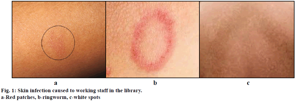
The aeromycoflora were collected from the indoor environment of two sections of selected library. One section comprises of reference section i.e. reading room and the other was stack section where books are stored in racks. The petri plate exposure culture plate method was employed for determination of total number of colonies recorded in the entire sampling period. Sterilized petri plates of 10 cm diameter containing potato dextrose agar (PDA) and Czapek Dox agar (CDA) media were exposed for 10 minutes. Streptomycin was added to inhibit bacterial growth on the media. The petri plates were incubated at 28±1° for 4-5 days. The colonies appeared on mother agar plates, each colony was inoculated on the separate petri plate containing PDA and CDA media. The petri plates were sealed with paraffin tape and were kept in incubator for 5-6 days at 28±1°. After full growth of the pure cultures, fungi were transferred to slants. The isolated fungi were examined and identified with the help of authentic literature. The staff working in the library was suffering from skin infections were observed during the research period.
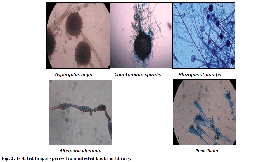
Isolation of fungi from infested or damaged and deteriorated samples of books in library was also made. The selected samples were categorized as books of different age and colours and isolation of fungi associated with books, was made by the cotton swab method using PDA and CDA media. Fungal isolates showed the different colours on deteriorated books (Table 1).
| Fungi isolated | Colours on infested books |
|---|---|
| A. niger | Black |
| P. chrysogenum | Blue-green |
| R. stolonifer | Dirty white |
| C. spiralis | Grey |
| A. alternata | Olive black or brown |
Table 1: Fungi Isolated from Infested Books and their Colours on Books
The isolated fungi from books in library were studied further for cellulolytic activity by weight loss test method after incubation for 10 and 15 days [17]. Five isolates of the cellulolytic fungal species namely, A. niger, Penicillium chrysogenum, Rhizopus stolonifer, Chaetomium spiralis, Alternaria alternata were screened for cellulose degrading activity. The methodology used was a modification of the technique described by Fergus [18]. The isolates were grown individually on Czapek Dox Broth, with newspaper and book paper as the sole source of carbon. The experiments were carried out in petri plates of 90 mm diameter. Petri plates contained newspaper and book paper discs of known weight along with 10 ml of Czapek Dox Broth without sucrose. Triplicates were maintained for monitoring the results on 10th and 15th d. Similar sets maintained in identical conditions were served as the control. All plates prepared in this manner were autoclaved at 15 lb psi for 20 min.
The inoculum was prepared in the form of a uniform suspension of spores (106 spores/ml) from 15 d old cultures of the respective isolates grown on CDA medium by adding 10 ml of sterile distilled water, followed by shaking on a vortex mixer. One milliliter of the suspension was added to each of the respective plates as inoculum. In the control set sterile distilled water was added instead of spore suspension. The plates were incubated at 28° for 10th and 15th d, respectively. At the end of the respective incubation periods newspaper and book paper discs were oven dried at 80°, allowed to cool down to ambient temperature in a desiccator and then weighed on a balance. The difference in weight of newspaper and book paper discs was computed by comparing it with the original dry weight and also of the control set. The net loss in weight was attributed to cellulose degradation. The percent loss in weight brought about by each isolate was calculated using the formula [19], % loss in weight=(difference in weight/ initial weight)×100. Temperature and relative humidity were recorded in the libraries during the sampling period using a hygrometer (Table 2).
| Months | Temperature | Humidity | ||||||
|---|---|---|---|---|---|---|---|---|
| R | S | R | S | |||||
| Max | Min | Max | Min | Max | Min | Max | Min | |
| May | 39.2 | 31.2 | 40.1 | 35.5 | 48 | 30 | 47 | 24 |
| June | 31.1 | 26.5 | 33.8 | 32.7 | 79 | 64 | 77 | 63 |
| July | 23.1 | 22.4 | 26.9 | 26.3 | 87 | 80 | 87 | 85 |
| August | 28.9 | 26.9 | 31.5 | 29.3 | 84 | 75 | 82 | 73 |
| September | 30.7 | 29.9 | 33.4 | 30.1 | 78 | 62 | 74 | 61 |
| October | 29.9 | 28.6 | 31.7 | 28.9 | 78 | 69 | 78 | 70 |
Table 2: Average Temperature and Relative Humidity Recorded from two Sections of Library
The mycoflora trapped from the air inside the college library were observed to be A. niger, A. fumigatus, A. flavus, A. caespitosus, A. alternata, A. tenuissima, Rhizopus stolonifer, Curvularia lunata, Chaetomium spiralis, P. chrysogenum, Fusarium pallidoroseum, Helminthosporium solani, Geotrichum candidum and Drechslera tetramera. Among the various species encountered A. niger and A. fumigatus were the dominant fungi inside the library and mostly these Aspergillus species were also responsible for the skin infections to the staff of library [20] analysed during the study period on the body parts of staff members as shown in Figure 1.
Out of the two sections of library, the occurrence of fungal species was examined more in the stack section than that in the reference section. It may be because the wet and humid conditions in the stack section induced the occurrence of mycoflora more than that of reference section. In both sections of the library, more concentration of total fungal species was contributed by the Aspergillus group (Table 3). Monthly variations in the total mycofloral concentration were observed during the study [21]. The concentration of mycoflora was recorded highest in the month of August, September and October than May, June and July. Out of the fungal species isolated from indoor air of library, some of the fungi like A. niger, P. chrysogenum, R. stolonifer, C. spiralis and A. alternata were also isolated from the infested books in library as shown in Figure 2.
| Fungal types | Library sections | May | June | July | Aug | Sep | Oct | Total species |
|---|---|---|---|---|---|---|---|---|
| A. niger | R | 12 | 10 | 08 | 05 | 02 | 0 | 37 |
| S | 18 | 16 | 07 | 09 | 02 | 03 | 55 | |
| A. fumigatus | R | 04 | - | 06 | - | 01 | 03 | 14 |
| S | - | - | 08 | 05 | 03 | 06 | 22 | |
| A. flavus | R | 02 | - | - | 04 | 01 | - | 07 |
| S | - | - | - | 05 | 02 | 01 | 08 | |
| A. caespitosus | R | - | - | - | 01 | 02 | 03 | 06 |
| S | 01 | - | - | 03 | 06 | 05 | 15 | |
| P.chrysogenum | R | - | 07 | - | 02 | - | 01 | 10 |
| S | - | 01 | 01 | 02 | - | 03 | 07 | |
| A.alternata | R | - | 04 | - | - | 03 | 06 | 13 |
| S | 02 | - | 01 | 02 | 09 | 08 | 22 | |
| A. tenuissima | R | - | 01 | - | - | - | 01 | 02 |
| S | - | - | 02 | - | 02 | 05 | 09 | |
| C. lunata | R | - | - | 01 | 04 | 01 | - | 06 |
| S | 02 | - | - | 02 | 02 | 05 | 11 | |
| F. pallidoroseum | R | - | - | - | - | 01 | 03 | 04 |
| S | - | - | - | 01 | 01 | - | 02 | |
| R. stolonifer | R | 01 | - | - | 03 | 02 | 01 | 07 |
| S | - | - | 03 | - | 04 | 06 | 13 | |
| C. spiralis | R | - | - | - | 02 | 01 | - | 03 |
| S | - | - | - | - | 02 | - | 02 | |
| H. solani | R | - | 01 | - | 01 | - | 01 | 03 |
| S | - | - | - | - | 02 | 03 | 05 | |
| G. candidum | R | - | - | - | 05 | - | - | 05 |
| S | 01 | - | - | 04 | 03 | - | 08 | |
| D. tetramera | R | - | 02 | - | 01 | - | - | 03 |
| S | - | - | - | - | 04 | - | 04 | |
| Unidentified fungi | R | 02 | 01 | 02 | 02 | 01 | 07 | 15 |
| S | 03 | 03 | 02 | 05 | 04 | 03 | 20 |
Table 3: Showing Occurrence of Fungal Species in the Two Sections ao a Library
Cellulolytic activity of some book-isolated fungi has been studied on newspaper and book paper. The cellulolytic activity was estimated by the weight loss test. A. niger and C. spiralis showed same percent loss of substrate i.e. 30% on 10th d on book paper. The weight loss on Newspaper on 10th d was about 29.70% caused by C. spiralis followed by A. niger, which was 18.90%. A. niger caused a high percent loss of weight (35%) on 15th d of book paper followed by C. spiralis, 34%, while C. spiralis caused a greater % loss of weight (32%) on 15th d of newspaper which is followed by A. niger 21.60%. Table 4 showed the percent loss in weight of the paper discs produced by the test fungi. From the results of this study, it could be concluded that the maximum cellulolytic activity in terms of loss in weight of both the papers was caused by A. niger and C. spiralis as compared to the other fungi tested. A. niger, C. spiralis and A. alternata affected books more than the newspaper and P. chrysogenum, R. stolonifer affected the newspaper more than books. The time course indicated that the degradation was less intensive especially at the first stages of infection i.e. on 10th d. However, on 15th d, an increase of degradation was observed but at a lower rate as maximum loss in weight occurred in the first 10 days of the experimentation period. The maximum cellulolytic activity recorded by the organisms in the initial period was apparently due to the abundant availability of moisture content in newspaper and book paper. The loss in weight of the papers indicated the amount of paper degraded by the fungus, thereby reflecting its cellulolytic ability.
| Fungi tested | % loss in NP | % loss in BP | ||
|---|---|---|---|---|
| Period of incubation | Period of incubation | |||
| 10 d | 15 d | 10 d | 15 d | |
| A. niger | 18.90 | 21.60 | 30.00 | 35.00 |
| P. chrysogenum | 17.50 | 20.00 | 14.20 | 15.90 |
| R. stolonifer | 16.10 | 18.80 | 12.80 | 14.50 |
| C. spiralis | 29.70 | 32.00 | 30.00 | 34.00 |
| A. alternata | 15.80 | 18.40 | 19.90 | 22.70 |
| 0.90 (+) | 0.90 (+) | 0.92 (+) | 0.92 (+) | |
Table 4: The Percentage Loss of Substrates by Some Book Deteriorating Fungi
Acknowledgments
Authors express their sincere gratitude to the Head of the Department of Botany, Hislop College, affiliated to R.T.M. Nagpur University, Nagpur for providing laboratory facilities.
Conflict of interest
No potential conflict of interest was reported by the authors.
Financial support and sponsorship
Nil.
References
- Meraj-ul-Haque, Bhowal M, Patil A. Diversity of aeromycoflora in indoor and outdoor environment.Imperial J Interdisciplinary Res 2016;2:240-8.
- Durugbo EU, Kajero AO, Omoregie EI, Oyejide NE. A survey of outdoor and indoor airborne fungal spora in the Redemption City, Ogun State, southwestern Nigeria. Aerobiologia 2013;29:201.
- Lacey J. The Aerobiology of conidial fungi. In: Cole GT, Kendrick B, editors. Biology of conidial fungi. New York: Academic Press; 1981. p. 373-416.
- Lehrer SB, Aukrush L, Salvaggio JE. Respiratory allergy induced by fungi. Clin Chest Med 1983;4:23-41.
- Ghosh D, Dhar P, Chakraborty T, Uddin N, Das AK. Study of aeromycoflora in indoor and outdoor environment of national library, Kolkata.Int J Plant Animal Environ Sci 2014;4:663-72.
- Kalbende S, Dalal L, Bhowal M. The monitoring of airborne mycoflora in the indoor air quality of library. J Nat Prod Plant Resour 2012;2:675-9.
- Dalal L, Bhowal M, Kalbende S. Incidence of deteriorating fungi in the air inside the college libraries of Wardha city. Arch ApplSci Res 2011;3:479-85.
- Vittal BPR, Glory AL. Airborne fungus spores of a library in India. Grana 1985;24:129-32.
- Tilak ST, Pillai SG. Fungi in Library: an aerobiological survey. Ind J Aerobiol 1988;1:92-4.
- Singh A, Ganguli M, Singh AB. Fungal spores are an important component of library air. Aerobiol 1995;11:231-7.
- Sinha A, Singh MK, Kumar R. Aerofungi-An important atmospheric biopollutant at atmosphere. Indian J Aerobiol 1998;11:19-23.
- Pavan R, Manjunath K. Indoor and outdoor air quality of poultry farm at Bangalore. Int J Pharm Bio Sci 2014;5:654-65.
- Pavan R, Manjunath K. Qualitative analysis of indoor and outdoor airborne fungi in cowshed. J Mycol 2014;2014:985921.
- Ajoudanifar H, Hedayati MT, Mayahi S, Khosravi A, Mousavi B. Volumetric assessment of airborne indoor and outdoor fungi at poultry and cattle houses in the Mazandaran Province, Iran.ArhHigRadaToksikol 2011;62:243-8.
- Afshari MA, Riazipour M, Kachuei R, Teimoori M, Einollahi B. A qualitative and quantitative study monitoring airborne fungal flora in the Kidney Transplant Unit. NephroUrol Mon 2013;5:737-40.
- Gofron AG, Bosiacka B. Effects of meteorological factors on the composition of selected fungal spores in the air. Aerobiol 2015;31:63-72.
- Kolet M. Quantitative analysis of cellulolytic activity of the genus Chaetomium-I. Bionano Frontier 2010;3:304-6.
- Reddy PLN, Babu BS, Radhaiah A, Sreeramulu A. Screening, identification of cellulolytic fungi from soils of Chittoor District, India. Int J Curr Microbiol App Sci 2014;3:761-71.
- Ghewande MP. Decomposition of cellulose and production of cellulolytic enzymes by pathogenic fungi. J Biol Sci 1977;20:69-73.
- Patel SI. Studies on Potential Aero Allergens in the College Libraries. OIIRJ 2015;5:53-7.
- Adhikari A. Investigations on aero-mycology in relation to allergy in some selected semi-rural places of West Bengal, India[Ph.D. dissertation].Calcutta:Jadavpur University; 2000.


