- *Corresponding Author:
- K. S. Nagarsekar
Departments of Pharmacognosy, Pharmaceutics, Bombay College of Pharmacy, Kalina, Mumbai-400 098, India
E-mail kalpanagarsekar@gmail.com
| Date of Submission | 4 January 2011 |
| Date of Revision | 25 July 2011 |
| Date of Acceptance | 1 August 2011 |
| Indian J Pharm Sci, 2011, 73 (4): 422-429 |
Abstract
Supercritical fluid extract and ethanol extract of Vitex negundo Linn. were subjected to the chromatographic evaluation for identification of their constituents. Free radical scavenging activity of both extracts was studied by subjecting them to DPPH assay. IC50 values of ethanol and supercritical fluid extract of Vitex negundo indicate that ethanol extract has stronger reducing potential and ability to scavenge free radicals as compared to the supercritical fluid extract. The in vivo effect of extracts on lipid peroxidation was studied using ethanol induced oxidative stress model in rat. Ingestion of extracts for 14 days exhibited significant reduction in plasma MDA level of stressed animals. Ethanol extract exhibited higher in vivo antilipid peroxidation potential as compared to supercritical fluid extract which correlated well with radical scavenging potential of extract.
Keywords
Antioxidant potential, oxidative stress, supercritical fluid extract, Vitex negundo
Nirgundi (Vitex negundo L.) is widely used in Ayurvedic medicine in India. Although all parts of Vitex negundo are used in the indigenous system of medicine, leaves are the most potent for medicinal use. Leaves are reported to possess significant antiinflammatory [1], anticonvulsant [2] and analgesic activity [3]. These activities may be attributed to high antioxidant potential of its constituents. Gupta et al. have reported polyphenolic compounds, terpenoids, glycosidic iridoids and alkaloids as important constituents of the leaves [4]. Presently, large amounts of environmentally unfriendly solvents and technologies operating with high costs are employed in extraction of these biologically active compounds. An alternative extraction technique with better selectivity and efficiency is highly desired in order to eliminate solvents, avoid the degradation or loss of sensitive substances and to decrease the high energy and manpower inputs of conventional processes. Supercritical fluid extraction (SFE) is an efficient and environmentally benign extraction technique for solid materials and has been extensively studied for the separation of active compounds from herbs and other plants.
The interest in SFE has increased in recent years because of legal limitations of solvent residues and solvents which makes the process more suitable in pharmaceutical industries [5]. Of all the gases and liquids studied, CO2 remains the most commonly used fluid for SFE applications because of its low critical parameters (Tc=31.04°, Pc=73.8 bar), its non-toxic and nonflammable properties and its availability in high purity at low cost. Supercritical CO2 has good solvent properties for extraction of non-polar components such as hydrocarbons [6]. Although the antioxidant properties of leaves of Vitex negundo are well known, no published literature exists about the properties of the extracts obtained by SFE. Consequently, the aim of this study was standardization of supercritical fluid extract of Nirgundi and its detailed evaluation for antioxidant potential in comparison with extract prepared by conventional maceration and percolation method.
Materials and Methods
The leaves of Vitex negundo were obtained from medicinal garden of Bombay College of Pharmacy, Mumbai, India. The leaf sample was authenticated by Agharkar Research Institute, Pune (Auth08- 013) as leaves of Vitex negundo Linn. family Verbenaceae. The shade dried and ground raw material was a greenish-brown powder with a characteristic odor. This was used for all the extractions. Moisture content of the dried powder was 12.87±0.62% (w/w).
The CO2 used was 99.5% (w/w) pure and supplied by Alchemie gases, India. Ethyl alcohol was used for conventional extraction by maceration and percolation method. Analytical grade reagents (s.d. fine chemicals Ltd., India) were used for chemical analysis.
Extraction
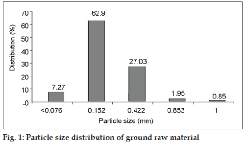
Previous studies revealed that the solvent consumption of SFE decreased significantly when the plant material was ground before the extraction [7]. Thus experiments were carried out using ground dried leaves. The particle size distribution of ground powder was analyzed by sieving with a set of sieves of different mesh sizes. A typical particle size distribution of dried powdered leaves used for supercritical fluid extraction is shown in fig. 1.
SFE was carried out in a high-pressure apparatus SFT-100 Supercritical Fluid Processing Unit (SFPU) (Supercritical fluid technology, Inc. Newark, U.S.A.), equipped with a 500 ml volume extractor vessel, using the supercritical fluid carbon dioxide. Samples of 200 g of the plant material were weighed accurately and put into the extraction vessel. The total extract of the plant material was recovered at 414 bar and 40°. The flow rate of CO2 was kept at 23.97 ml/min, density at working pressure and temperature was 0.896 g/cm3 and the extraction was carried out for 201 min. The extract was collected in externally mounted sample vial. The yield of SFE was 0.882 g/100 g dry material.
The powdered dried leaves were extracted using 96% ethanol (Herb:menstrum ratio 1:8) by maceration in stoppered container with frequent agitation for 48 h. The filtrate was separated, extracted leaves were further kept for percolation for 3 days and subsequently the extraction procedure was repeated until the percolate was almost colorless. The extract was combined and solvent was evaporated in vacuum (0.33 bar) in a rotary evaporator (Buchi, Germany) at 55-60°. The yield of ethanol extraction was 15.73% w/w.
Evaluation of extracts for presence of flavonoids by chromatography
One thousand parts per million solution of ethanol extract and supercritical fluid extract were prepared and sonicated on bath sonicator (Expo High-tech). Ten microlitres of each solution was applied in triplicate on a precoated aluminum (10×10 cm2) TLC plate (silica gel 60 F254, E. Merck) of uniform thickness of 0.2 mm. The plates were then conditioned for 20 min in a pre-saturated twin-trough chamber (Camag, Muttenz, Switzerland, 10×10 cm2) in solvent system ethyl acetate:formicacid:acetic acid:water (100:11:11:27 v/v/v/v) [8]. After development to a distance of 8 cm, plates were air-dried. The visualization of flavonoids and phenolic acids was achieved by spraying the plates with NP/PEG reagent. Plate was observed under 365 nm in UV-light.
Qualitative analysis of negundoside in ethanolic extract by HPTLC
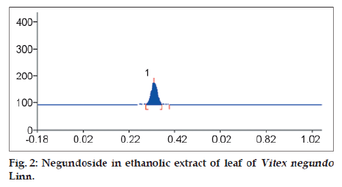
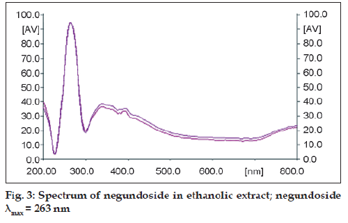
Presence of negundoside in the ethanolic extract was confirmed using TLC technique. Five microlitres of test solution of ethanolic extract (500 ng/ml) was applied on a precoated aluminum (10×10 cm2) TLC plate (silica gel 60 F254, E. Merck) of uniform thickness of 0.2 mm (n=3). The plates were then conditioned for 20 min in a pre-saturated twin-trough chamber (Camag, Muttenz, Switzerland, 10×10 cm2) in solvent system ethyl acetate:water:acetic acid (8:1:1 v/v/v) and developed to a distance of 8 cm. After development, plates were air dried and densitometric scanning was then performed at a wavelength of 260 nm with Camag TLC scanner III operated in the reflectance-absorbance mode and controlled by WinCATS software (v1.2.1). The slit dimension was kept at 5 mm×0.45 mm and scanning speed employed was 20 mm s−1. The spot for negundoside was confirmed by comparing the colour, Rf and spectrum of the spot with that reported in literature for pure negundoside (fig. 2). To ascertain the presence of negundoside in the extract the spot at Rf 0.35 in all tracts was subjected to spectrum scan from 200-800 nm [4] (fig. 3).
Preliminary TLC study for evaluation of volatile oil components
Thin layer chromatographic technique was used for preliminary evaluation of volatile oil components in extracts of Nirgundi. 2 mg/ml of test solution of supercritical fluid extract and ethanol extract in chloroform was prepared by sonicating mixture in bath sonicator. 5 μl of each test solution was applied in triplicate on a precoated aluminum (10×10 cm2) TLC plate (silica gel 60 F254, E. Merck). The plates were then conditioned for 20 min in a pre-saturated twin-trough chamber (Camag, Muttenz, Switzerland, 10×10 cm2) in solvent system toluene:ethyl acetate 93:7 v/v [9]. After development to a distance of 8 cm, plates were air dried. The visualization of constituents on plate was achieved by spraying plate with anisaldehyde/sulphuric acid reagent and heating plate at 100° for 10 min then evaluated in visible light.
GC/MS analyses of supercritical fluid extract
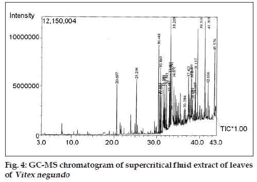
Presence of phytoconstituents in supercritical fluid extract was evaluated using GC/MS analysis. The solution of supercritical fluid extract in chloroform was analyzed using GC–MS unit which consisted of a Shimadzu GC 2010 gas chromatograph (Japan) equipped with DB-5 MS. fused-silica column (30 m×0.25 mm i.d., film thickness 0.25 μm; J and W Scientific Inc., Agilent Technologies, Santa Clara, CA, USA), and interfaced with QP 2010 quadrupole mass analyser. Oven temperature was programmed, 60° for three mins, subsequently at 5°/min up to 160°, held isothermal for 2 min; then raised upto 300° with 10° and held isothermal. Injector and detector temperatures were maintained at 250° and 280°, respectively. The eluted peaks were integrated and identified using NIST library search method (fig. 4).
Characterization of supercritical fluid extract by quantification of β-caryophyllene using GC analysis
Supercritical fluid extract of Nirgundi was characterized using β-caryophyllene as standard. Standard solution of β-caryophyllene (2 mg/ml) was prepared in N,N-dimethyl formamide (DMF). The solution was diluted suitably to give solutions containing β-caryophyllene in concentration range of 0.25 mg/ml to 1 mg/ml. The sample solution of supercritical fluid extract (12.4 mg/ml) was prepared in DMF. Quantitative analysis of 2 ml of standard solutions and sample solution were performed in a Varian CP-3800 gas chromatograph (Germany), equipped with autosampler with headspace and a flame ionization detector (FID). The analysis was carried out with the conditions given in Table 1. Standard plot obtained by plotting concentration of β-caryophyllene in mg against peak areas. β-caryophyllene in SCF was calculated and compared with that in hydrodistilled oil prepared by hydrodistillation using Clevenger’s Apparatus. Fig. 5 represents gas chromatograms of standard β-caryophyllene and supercritical fluid extract.
| Parameters | Conditions |
|---|---|
| Column | Fused silica capillary column (0.25 mm×30 m) with 0.25 μm coating of free fatty acid phase |
| Oven temperature | Programmed from 90° to 210° at 7°/min |
| Injector temperature | 230° |
| Syringe temperature | 90° |
| Sample equilibrium temperature | 80° |
| Detector temperature | 240° |
| Carrier gas | Nitrogen, hydrogen, air |
| Flow rate | 1.5 ml/min |
| Injector mode | Head space |
Table 1: Gc Conditions
Free radical scavenging activity of extracts (In vitro antioxidant assay)
DPPH is most common stable radical commonly used in antioxidant assays. Free radical scavenging activity of solutions of supercritical fluid extract and ethanol extract of leaves of Vitex negundo Linn. was assessed using DPPH radical. Different concentrations of test agents were prepared to find effective concentration or EC50 value. Decrease in optical density of the solution was read at 520 nm. The IC50 values of both test agents were determined. Free radical scavenging activity was calculated on the basis of % inhibition [10]. The formula for, % inhibition=(Acontrol)−(Atest)/(Acontrol)×100.
Effect of extracts on oxidative stress (In vivo antioxidant model)
The objective of the study was to investigate effect of drug extracts on oxidative stress induced in rats by administration of alcohol. Females of Sprague dawley strain (150-180 g) were used. Animals were fed and housed as per standard guidelines of CPCSEA. The animals were given a week time to get acclimatized to the laboratory conditions. The protocol was approved by the institutional ethics committee.
Animals were divided into four groups of six animals each. Group I served as disease control in which 1 ml/kg/d of 30% v/v ethanol was instilled intragastrically for 15 days. Group II received dose of 500 mg/kg/d of ethanolic extract of leaves of Vitex negundo Linn. in 0.5% CMC and 30% v/v ethanol, intra-gastrically for 15 days. Group III received dose of 500 mg/kg/d of supercritical fluid extract of leaves of Vitex negundo Linn. in 0.5% CMC and 30% v/v ethanol, intra-gastrically for 15 days. Group IV served as vehicle control in which 1 ml/ kg/d of distilled water was instilled intra-gastrically for 15 days. At the end of treatment, 1-2 ml blood of each animal was collected from the inner canthus of the eye by retroorbital puncture, using capillary tube, under light ether anesthesia, in micro centrifuge tubes (2 ml) containing sodium citrate solution USP (4%) as anticoagulant. Plasma was separated by centrifugation at 4000 rpm for 10 min and was stored at −20°.
The estimation of malondialdehyde (MDA) was carried out by TBARS method. Thiobarbituric acid (TBA) reacts with MDA, one of the aldehyde products of lipid peroxidation, to give a colored product which was extracted in butanol and absorbance measured spectrophotometrically at 532 nm. 10 ml stock solution of MDA was prepared by adding 1 mmol of 1,1,3,3-Tetraethoxypropane (Sigma chemicals) to 100 ml of (1% v/v) sulphuric acid. The mixture was left at room temperature at 2 h to achieve complete hydrolysis. This stock was further diluted to about 10-100 μM. The absorbance of the solution was taken at 245 nm. 0.1 ml of MDA solutions of different concentrations were mixed with 1.5 ml of 20% acetic acid, 0.2 ml of 8.1% sodium lauryl sulphate and 1.5 ml of 0.8% TBA solution. The volume was adjusted to 4 ml with distilled water and the mixture was incubated at 100° for 60 min. The mixture was then cooled under the tap water. After cooling, 4 ml of n-butanol was added to it. The mixture was centrifuged at 4000 rpm for 10 min the absorbance of organic layer (upper layer) was read at 532 nm [11].
The standard curve was prepared correlating the concentration of malondialdehyde in solutions used and the absorbance of the coloured complex obtained by reaction with TBA.
Results and Discussion
The TLC separation of ethanol extract of Vitex negundo Linn. revealed presence of flavonoids. Typical intense yellow fluorescence in UV–light at 365 nm was observed at RF values 0.11, 0.15, 0.43, 0.57, 0.65 and at application position. Red orange fluroscent zones at solvent front are due to chlorophyll derivatives. While the green fluorescent zones were observed at Rf values 0.27 and 0.76. The yellow green fluorescent bands might be due to presence of Vitexin [8]. The supercritical fluid extract did not show presence of any flavonoid.
Chromatogram after separation of ethanol extract using solvent system ethyl acetate:water:acetic acid (8:1:1 v/v/v) showed zone of negundoside at Rf 0.35 in all the tracks. Zone of negundoside showed good resolution from zones of other constituents. Spectrum of negundoside showed λmax at 263 nm which confirmed presence of negundoside in ethanolic extract.
As the result of preliminary TLC study of Supercritical fluid extract of Vitex negundo Linn. leaves, after derivatisation with anisaldehyde/sulphuric acid reagent, 13 coloured zones were observed in visible light. Chromatogram of ethanol extract did not show presence of coloured zones after treatment with anisaldehyde/sulphuric acid reagent. The TLC profile of supercritical fluid extract showed presence of phenyl propane derivatives which cause quenching of fluorescence at 254 nm. Almost all components showed blue fluorescence on exposure to 365 nm. Detection with anisaldehyde/sulphuric acid reagent revealed violet zone at Rf 0.91 near solvent front which could be attributed to presence of β-caryophyllene. The pink violet zone at Rf 0.43 may be due to presence of caryophyllene oxide. The zones between Rf 0.5 to 0.65 might be due to terpene aldehydes or terpene esters. Red violet zones of constituents between Rf 0.1-0.4 complied with Rf values of terpene alcohols [9]. Major constituents of leaf extracts are listed in Table 2.
| Extract | Main constituents | Calculated retention indices | Bibliographic retention indices | References |
|---|---|---|---|---|
| Ethanol extract | Negundoside | 0.35 | 0.35 | 4 |
| Supercritical fluid extract | β-carryophylline | 0.91 | 0.9 | 9 |
Table 2: Main constituents of leaf extracts Of Vitex negundo
The GC-MS analysis of supercritical fluid extract of Vitex negundo Linn. confirmed presence of β-caryophyllene (4.59%) and epiglobulol (6.60%) as major constituents. In addition esters of hexadecanoic acid, octadecanoic acid, linolenic acid and aliphatic hydrocarbons were found in the supercritical fluid extract.
The composition of the supercritical fluid extract of Vitex negundo Linn. was found to be significantly different than that of hydrodistilled essential oil which was reported to contain 4-terpineol, (z)-β- farnesine and caryophyllene oxide in considerable quantity. Amount of β-caryophyllene obtained from supercritical fluid extraction is significantly higher than that obtained by hydrodistillation method [12]. This can be attributed to low operating temperature used in SFE which reduce thermal degradation of most of the labile compounds. In addition the extract is free from solvent residue and extraction requires shorter extraction period.
In order to quantify the β-caryophyllene in SCFE, GC chromatograms of solutions of known concentration of standard β-caryophyllene were recorded. The plot of β-caryophyllene in mg vs. peak area was linear in the range of 0.25 mg to 2 mg as indicated by r2 value of 0.9981. The equation of the standard plot was as given as; y=25668x+1575, Where, y=peak area and x=β-caryophyllene in mg
The ammount of β-caryophyllene in SCFE obtained from 100 g of powdered leaves was found to be 40.4 mg which is significantly higher than that obtained by hydrodistillation method (5.4 mg/100 g of powdered leaves).
Free radical scavenging activity of solutions of Supercritical fluid extract and ethanol extract of leaves of Vitex negundo Linn. and their corresponding concentrations are as represented in Table 3. Fig. 6 indicate free radical scavenging activity of supercritical fluid extract and ethanolic extract.
| Conc. in μg/ml | Supercritical fluid extract | Ethanol extract | ||
|---|---|---|---|---|
| Mean absorbance | % Inhibition ± sd | Mean absorbance | % Inhibition ± sd | |
| DPPH control | 0.80 | – | 1.01 | – |
| 10 | 0.77 | 2.98 ± 0.03 | 0.96 | 5.12 ± 0.02 |
| 20 | 0.77 | 3.70 ± 0.03 | 0.94 | 7.25 ± 0.01 |
| 40 | 0.75 | 6.03 ± 0.01 | 0.86 | 15.03 ± 0.01 |
| 80 | 0.73 | 9.04 ± 0.03 | 0.75 | 25.52 ± 0.02 |
| 160 | 0.67 | 15.35 ± 0.02 | 0.57 | 43.70 ± 0.01 |
| 320 | 0.58 | 26.89 ± 0.02 | 0.23 | 77.16 ± 0.02 |
| 500 | 0.47 | 40.27 ± 0.02 | 0.17 | 82.66 ± 0.02 |
| 640 | 0.40 | 49.16 ± 0.03 | 0.15 | 84.91 ± 0.02 |
| 1000 | 0.31 | 61.37 ± 0.04 | 0.15 | 84.83 ± 0.03 |
| 1280 | 0.29 | 62.98 ± 0.02 | 0.15 | 84.98 ± 0.02 |
Table 3: free radical scavenging activity Of supercritical fluid extract and ethanol Extract
IC50 value of ethanol extract in DPPH assay was 190 μg/ml and the highest donor capability was achieved with 500 μg/ml of ethanolic extract (i.e. approximately 82.67% DPPH reduced) after which plateau effect was observed. Supercritical fluid extract showed IC50 value for 670 μg/ml and the highest donor capability was achieved with 1000 μg/ml (i.e. approximately 61.38% DPPH reduced) after which plateau effect was observed. The degree of discolouration indicates scavenging capacity of the extract. The IC50 values of extracts indicate that Ethanolic extract has stronger reducing potential and ability to scavenge free radicals as compared to the supercritical fluid extract.
In recent years oxidative stress has been implicated in pathophysiology of many diseases or disorders which are initiated or exacerbated by pro-oxidants such as various drugs, alcohol and food additives etc [13]. Ethanol-induced oxidative stress model is reported in literature for evaluation ability of plant extract to reduce the oxidative stress [14]. Ethanol is almost exclusively metabolized in the body by enzyme catalyzed oxidative processes. Drinking high doses of alcohol for longer period of times increases activity of enzyme called cytochrome P450 2E1. This triggers cascade of events which cause liver injury [15]. Injested alcohol produces striking metabolic imbalance in liver which leads to formation of reactive oxygen species (ROS). Inadequate removal of the ROS may cause cell damage by attacking membrane lipids, proteins and inactivating enzymes, thus mediating several forms of tissue damage [14]. Lipid peroxidation of polyunsaturated fatty acids in membranes has been implicated as a mechanism by which ethanol causes liver damage [16]. ROS react with fatty acids leading to accumulation of lipid peroxidation products which include malondialdehyde. This compound is most commonly used as a marker of the degree of lipid peroxidation [17]. The antioxidants seize the free radical chain of oxidation and form a stable endpoint, which do not permit further oxidation of lipid.
The current study evaluated the effect of Vitex negundo Linn. extract on plasma lipid profile in ethanol-administered rats. The amount of MDA formed is quantified by reaction with thiobarbituric acid (TBA). Actual concentration of MDA is found using the molecular extinction coefficient. Concentration of MDA=Absorbance at 254 nm/ε, where ε is molecular extinction coefficient [11].
As represented in Table 4 MDA level of the rats of vehicle control group i.e. alcohol untreated was fairly constant to the value of 2.7 nmol/ml. In the study presented administration of 30% ethanol 1 ml/ kg/d caused increase in level of MDA up to 4.37 nmol/ ml in rats of positive control group confirm the oxidative stress in SD female rats. Stressed animals when treated with 500 mg/kg/d of ethanolic extract for 14 days exhibited significant reduction in MDA level to the value 2.89 nmol/ml. This is statistically equivalent to the mean MDA value in control group animals when subjected to students t-test (P>0.05). This suggested utility of ethanol extract in reducing lipid peroxidation and MDA concentration to normal levels. Whereas MDA level of supercritical extract treated stressed animals was found to be 3.46 nmol/ml which is higher than the levels of normal control but subsequently lower than the MDA levels in positive control group (P<0.05).
| Group | no Drug | Dose (mg,ml/kg p.o./ daily×15 d | MDA levels in n mol/ml |
|---|---|---|---|
| I | Ethanol (disease control) | 1 | 4.36 ± 1.98 |
| II | VNE+Ethanol | 500+1 | 2.88 ± 1.29 |
| III | SCFE+Ethanol | 500+1 | 3.46 ± 2.30 |
| IV | Vehicle control (DW+CMC) | 10 | 2.70 ± 2.19 |
Table 4: The Mda levels of plasma after the Extract treatment
The leaves of Vitex negundo Linn. are known to posses various antioxidant chemical constituents which are relatively polar in nature like flavonoids, carotene, vitamin C etc. which possibly be responsible for the reduction of lipid peroxidation in present study. The higher antioxidant potential of the extract may be due to the extraction of these constituents in ethanol which is a polar solvent (polarity 5.1).
The constituents in supercritical fluid extract are relatively nonpolar. As per preliminary chromatographic test results and Gas-chromatography mass-spectrometric analysis these constituents are terpens and terpenoidal compounds. These constituents in supercritical fluid extract may possibly be responsible for its antioxidant and antilipid peroxidation potential. Many constituents have phenolic hydroxyl groups that can act as hydrogen donors and hence posses antioxidant potential.
The antilipid peroxidative property of the extract is expected to relate to its radical scavenging potential. Hence protection offered by both the extracts against lipid peroxidation can be related to their antioxidant nature.
In conclusion, essential oils were extracted from leaf powder of Vitex negundo Linn. by supercritical fluid extraction. TLC fingerprints of these extracts suggested presence of non polar constituents viz. terpenes which was confirmed by GC and GC-MS analysis. Ethanol extract of leaves showed presence of polar constituents like flavonoids in TLC. Both the extracts exhibited radical scavenging ability in in vitro testing and this was in correlation with reduced levels of TBARS when extracts were orally administered to rats, indicating usefulness of the extracts in reducing lipid peroxidation. Antilipid peroxidation potential of leaves of Vitex negundo is due to radical scavenging potential of both polar and nonpolar constituents. Vitex negundo Linn. leaf extracts have potential to be drug/adjunct in management of diseases caused by oxidative stress.
Acknowledgements
Authors are thankful to Mr. C. R. Barhate, for help in preparation of manuscript, Mrs. Vaishali (M.K.R. Labs) for GC analysis, Anchrom Labs (Mulund) for chromatographic facilities.
References
- Tiwari OP, Tripathi YB. Antioxidant properties of different fractions of VitexnegundoLinn. Food Chem 2007;100:1170-6.
- Tandon VR, Gupta RK. An experimental evaluation of anticonvulsant activity of Vitexnegundo. Indian J PhysiolPharmacol 2005;49: 199-205.
- Gupta RK, Tandon VR. Antinociceptive activity of Vitexnegundo Linn. leaf extract. Indian J PhysiolPharmacol 2005;49:163-70.
- Gupta A, Tandon N, Sharma M. Vitexnegundo Linn. Quality standards of Indian medicinal plants. Vol. 6, New Delhi: Indian Council of Medical Research; 2008. p.357-66.
- Vági E, Simándi B, Suhajda Á, Héthelyi É. Essential oil composition and antimicrobial activity of Origanummajorana L. extracts obtained with ethyl alcohol and supercritical carbon dioxide. Food Res Int 2005;38:51-7.
- Qingyong L, Chien MW. Supercritical fluid extraction in herbal and natural product studies- a practical review. Talanta 2001;53:771-82.
- Simándi B, KristoSzT, Kéry Á, Selmeczi LK, Kmecz I, Kemény S. Supercritical fluid extraction of dandelion leaves. J Supercrit Fluids 2002; 23:135-42.
- Flavonoid Drugs including gingko biloba. In: Wagner H, Bladt S, editor. In Plant Drug Analysis A thin layer chromatography atlas. 2nd ed. Germany: Spriner-Verlag; 1996. p. 195-244.
- Drugs containing Essential oils, balsams, oleo gum resins. In: Wagner H, Bladt S, editors. Plant Drug Analysis A thin layer chromatography atlas. 2nd ed. Germany: Spriner-Verlag; 1996. p. 149-92.
- Baydar N, Ozkan G, Yasar S. Evaluation of antiradical and antioxidant potential of grape extracts. Food Control 2007;18:1131-6.
- Esterbauer H, Cheseman K. Determination of aldehydic lipid peroxidation products: Malondialdehyde and 4-hydroxynonenal. Meth Enzymol 1990;186:407-21.
- Nagarsekar KS, Nagarsenker MS, Kulkarni SR. Evaluation of composition and antimicrobial activity of supercritical fluid extract of leaves of Vitexnegundo. Indian J Pharm Sci 2010; 72:641-3.
- Gupta S, Pandey R, Katyal R, Aggarwal S. Lipid peroxide levels and antioxidant status in alcoholic liver disease. Indian J ClinBiochem 2005;20:67-71.
- Gupta R, Tandon V. Effect of Vitexnegundo on oxidative stress. Indian J Pharmacol 2005;37:38-40.
- Nanji A, French S. Animal models on alcoholic liver disease: Focus on intragastric feeding model. Alcohol Res 2003;27:325-30.
- Stal P, Johansson II, Hagen K, Hulkcrantz R. Hepatotoxicity induced by iron overload and alcohol. J Hepatol 1996;25:538-46.
- Purohit V, Russo D, Coater P. Role of fatty liver, dietary fatty acid supplements and obesity in progression of alcoholic liver disease: Introduction and summery of symposium. Alcohol 2004;34:3-8.

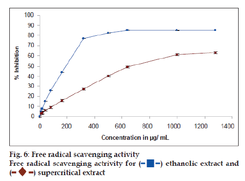
 ethanolic extract and
 supercritical extract


