- *Corresponding Author:
- Hanyi Li
Department of Gynecology and Obstetrics, Ganxi Cancer Hospital, Pingxiang, Jiangxi Province 337000, China
E-mail: 870827564@qq.com
| Date of Received | 19 December 2022 |
| Date of Revision | 12 May 2023 |
| Date of Acceptance | 06 November 2023 |
| Indian J Pharm Sci 2023;85(6):1816-1820 |
This is an open access article distributed under the terms of the Creative Commons Attribution-NonCommercial-ShareAlike 3.0 License, which allows others to remix, tweak, and build upon the work non-commercially, as long as the author is credited and the new creations are licensed under the identical terms
Abstract
The objective of this study is to examine the impact of microRNA-320 on the phosphoinositide 3-kinase/protein kinase B signaling pathway in ovarian tissue and serum fasting serum insulin levels and homeostasis model assessment of insulin in rats with polycystic ovary syndrome. A total of forty-eight female Westar rats, characterized by their clean-grade health status, were allocated into three groups; Control, model, and microRNA-320 overexpression. Each group consisted of sixteen rats. In comparison to the control group, the model group exhibited elevated levels of fasting serum insulin levels, fasting blood glucose, homeostasis model assessment of insulin, testosterone, estradiol, and luteinizing hormone, while the level of follicle-stimulating hormone was reduced. Conversely, when compared to the model group, the microRNA-320 overexpression group demonstrated increased serum levels of testosterone, estradiol, and luteinizing hormone, accompanied by a decrease in follicle-stimulating hormone level. The microRNA-320 overexpression group exhibited a significantly greater quantity of small follicles and atresia follicles compared to the model group, while the presence of granular cell layer and corpus luteum was not observed. In comparison to the control group, the expression levels of phosphoinositide 3-kinase, protein kinase B, glucose transporter 4, forkhead box O3, and p27 in the ovarian tissue of the model group exhibited a significant decrease. Furthermore, when compared to the model group, the expression levels of phosphoinositide 3-kinase, protein kinase B, glucose transporter 4, forkhead box O3, and p27 in the ovarian tissue of the microRNA-320 overexpression group also demonstrated a significant decrease. Overexpression of microRNA-320 can significantly inhibit the expression of phosphoinositide 3-kinase/protein kinase B signaling pathway protein in ovarian tissue of polycystic ovary syndrome rats, and up-regulate the serum levels of fasting serum insulin levels and homeostasis model assessment of insulin.
Keywords
microRNA-320, polycystic ovary syndrome, phosphoinositide 3-kinase/protein kinase B signaling pathway, fasting serum insulin levels, homeostasis, insulin
Polycystic Ovary Syndrome (PCOS) is a prevalent endocrine disorder syndrome characterized by the simultaneous presence of reproductive dysfunction and abnormal glucose metabolism. It represents the most frequently encountered endocrine and metabolic disorder among women in their childbearing years, and it also stands as a significant contributor to female infertility. The main manifestations are persistent anovulation, androgen excess and Insulin Resistance (IR)[1]. According to statistics, among women of childbearing age worldwide, the incidence rate is about 5 % to 10 %. The pathogenesis of PCOS is complex and unclear, which may be caused by the interaction between genetic and environmental factors[2,3]. microRNAs (miRNAs) can bind to the non-coding region of the target messenger Ribonucleic Acid (mRNA) 3 and further mediate the degradation, translation inhibition and deacetylation of the target mRNA. Some studies have found that miRNAs are abundantly expressed in the ovary, uterus, fallopian tube and other organs. miR-320, a member of the miRNAs family, participates in the regulation of insulin sensitivity in adipose tissue and is closely related to the occurrence and development of IR in patients with PCOS[4]. Furthermore, the Phosphoinositide 3-Kinase/Protein Kinase B (PI3K/Akt) signaling pathway assumes a significant role in the IR associated with PCOS[5]. However, the relationship between miR-320 to PI3K/Akt signal pathway and IR is not clear. The primary objective of this study was to examine the impact of miR-320 on the expression of PI3K/Akt signal pathway protein in ovarian tissue, as well as the levels of Fasting Insulin (FINS) and Homeostasis Model Assessment of IR (HOMA-IR) in the serum of rats with PCOS. Forty-eight clean-grade healthy female Wistar rats were randomly selected [Beijing Weitong Lihua, production license SCXK (Beijing) 2019-0009, use license (Beijing) 2017-0022]. They were 2 mo old and weighed (225.86±21.63) g. All rats were fed adaptively for 1 w at a laboratory temperature of 21°±2°, and humidity of 52 %±8 %, day and night for 12 h. Biological microscope (Shanghai Yuguang Instrument Factory, model: WMS-1030); tissue embedding machine (Shanghai Kehuai Instrument Co., Ltd., model: BMJ-1B); paraffin slicer (Jinhua Huiyou Instrument and equipment Co., Ltd., model: HY-1508). -80° cryogenic refrigerator (Nanjing Beidan Medical Co., Ltd., model: MDF-C8V); phosphate buffer (Jiangsu Enmoasai Biotechnology Co., Ltd.); Hematoxylin and Eosin (H&E) staining kit (Solebo Technology Co., Ltd.); PI3K antibody (Wuhan Boot Biotechnology Co., Ltd.); Akt antibody (Biyuntian Biotechnology Research Institute). Letrozole (Jiangsu Hengrui Pharmaceutical Co., Ltd., batch number: 20181001, specification: 2.5 mg×10 s). Rats were randomly divided into the control group, the model group and miR-320 overexpression group (n=16). Among them, the model group and miR-320 overexpression group; futurozole was dissolved into 1 % carboxymethyl cellulose according to 1 mg/kg/d and continuously fed for 30 d, while the control group of rats received solely a uniform dosage of 1 % carboxymethyl cellulose., and rats in the over-expression group were given 2 mg/kg miR-320 analogues through tail vein for 30 d. In the process of intragastric administration, the head and body of rats are at the same level. The rats were anesthetized on the experimental table. After successful anesthesia, the blood 5 ml of the abdominal aorta was taken and centrifuged at the speed of 3000 r/min. The concentrations of serum FINS and Fasting Blood Glucose (FBG) were measured using radioimmunoassay, and the HOMA-IR index was compute.
HOMA-IR=FBG (mmol/l)×FINS (mIU/l)/22.5
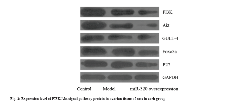
The levels of serum Testosterone (T), Estradiol (E2), Follicle-Stimulating Hormone (FSH) and Luteinizing Hormone (LH) were determined by Enzyme-Linked Immunoassay (ELISA). After blood collection, the rats were killed, exposed and bilateral ovaries were completely removed, and the surrounding adipose tissue was cut. The ovarian tissue was fixed with a 4 % formaldehyde solution, paraffin sections were routinely made and stained with H&E staining, and the histopathological changes of the ovary were observed under a microscope. The Western blotting technique was employed to ascertain the presence of miR-320 signal pathway protein expression in the ovarian tissues of rats belonging to the control group, model group and PI3K/Akt overexpression group. In this study, the counting data were compared by Chi-square (χ²) comparison, expressed by n (%). The independent sample t-test was employed to compare the measurement data between the groups, which was expressed as (x̄±s). In this study, Statistical Package for the Social Sciences (SPSS) 21.0 software was used to analyze the statistical data, the observed discrepancy was deemed to have statistical significance. In comparison to the control group, the model group exhibited elevated levels of FINS, FBG, and HOMA-IR. Furthermore, the miR-320 overexpression group demonstrated even higher levels of these variables compared to the model group. However, no significant disparity was observed in the FBG level between the two groups as shown in Table 1. In comparison to the control group, the model group exhibited elevated levels of serum T, E2 and LH, while the level of FSH was reduced. Conversely, the miR-320 overexpression group demonstrated increased levels of serum T, E2, LH and FSH in relation to the model group, albeit still lower than those observed in the model group as shown in Table 2. There are many layers of granulosa cells in the follicles of rats in the control group, and the presence of the corpus luteum is clearly evident, whereas in the model group, there is a scarcity of corpus luteum and follicles across all stages of development, the layers of granulosa cells in the follicles decrease or even disappear, and typical polycystic changes can be seen in the ovary; the small follicles and atretic follicles in the miR-320 overexpression group is more than that in the model group, and no obvious granulosa cell layer and corpus luteum were found as shown in fig. 1. In comparison to the control group, the model group exhibited decreased expression levels of PI3K, Akt, Glucose Transporter Type 4 (GLUT4), Forkhead box O3 (FOXO3), and P27 in the ovarian tissue. Furthermore, the ovarian tissue of the miR-320 overexpression group demonstrated even lower expression levels of these proteins in comparison to the model group as shown in fig. 2. PCOS is a prevalent and multifaceted condition observed among women within the reproductive age group, which is characterized by obesity, hyperandrogenemia, IR, chronic anovulation, hirsutism and acne. Studies have found that most patients with PCOS have a certain degree of abdominal obesity, which is closely related to the pathological characteristics of androgen excess, IR and inflammation[6]. As a result, the risk of patients with Type 2 Diabetes Mellitus (T2DM) and cardiovascular disease is significantly increased. With the development of medical science and technology, there are many ways to treat and improve the disease in clinics. Lifestyle changes and the application of insulin-sensitizing drugs are the most effective methods, but the specific mechanism is still unclear[7]. Therefore, the study on the pathology and drug intervention mechanism of PCOS has aroused the interest of scholars at home and abroad. miRNAs are a class of endogenous non-coding single-stranded RNA molecules that exert regulatory control over gene expression subsequent to transcription, thereby assuming a significant role in cellular processes such as growth and apoptosis. In recent years, some studies have found that miRNAs can regulate gene expression through targeting, thus participating in the occurrence and development of diseases[8]. Xiong et al.[9] confirmed that there are abnormal expressions of many types of miRNA in human ovaries, such as miR-143-3p, miR-29a, miR-26a, let-7 and so on, which have extensive effects on the regulation of ovarian function in many aspects. Studies have shown that miRNA may participate in the regulation and play a significant role in the progression of PCOS[10]. IR is characterized by a cascade of physiological and pathological alterations, wherein the metabolism and transportation of glucose in the human body are diminished below the standard physiological threshold due to the regulatory influence of insulin, that is, human target organs, tissues and cells are destroyed, weakened or lost by insulin, resulting in a significant decrease in absorption and glucose utilization efficiency[11]. To maintain a relatively normal level of blood glucose, insulin increases compensatively in the body, resulting in hyperinsulinemia. Its occurrence mechanism is more complex. The insulin signal transduction pathway includes the PI3K/Akt signal pathway and MAPK signal pathway.
| Group | FINS (mU/l) | FBG (mmol/l) | HOMA-IR |
|---|---|---|---|
| Control | 18.59±2.20 | 4.68±0.37 | 3.87±0.49 |
| Model | 30.94±4.07 | 5.88±0.84 | 8.96±1.09 |
| miR-320 overexpression | 38.94±4.08 | 5.98±0.92 | 10.35±1.13 |
| F | 132.59 | 14.87 | 206.52 |
| p | <0.001 | <0.001 | <0.001 |
Table 1: Serum FINS, HOMA-IR and FBG levels in rats (x̄±s, n=16).
| Group | T (ng/ml) | E2 (pg/ml) | FSH (mIU/ml) | LH (mIU/ml) |
|---|---|---|---|---|
| Control | 0.66±0.15 | 103.74±29.36 | 5.47±1.66 | 0.89±0.36 |
| Model | 1.89±1.42 | 126.78±20.19 | 3.56±1.10 | 6.48±1.46 |
| miR-320 overexpression | 4.09±2.42 | 145.06±22.92 | 2.52±1.26 | 12.89±6.15 |
| F | 18.36 | 11.46 | 19.35 | 43.18 |
| p | <0.001 | <0.001 | <0.001 | <0.001 |
Table 2: Comparison of serum T, E2, FSH and LH levels in rats of each group (x̄±s, n=16).
Among them, the PI3K/Akt signal pathway is mainly involved in the regulation of glucose metabolism in glucose-insulin post-receptor signal transduction, while the MAPK signaling pathway primarily participates in the modulation of cellular growth and differentiation[12]. It has been found that the PI3K/Akt signal pathway is the key to insulin post-receptor signal transduction[13]. IR caused by PCOS has an important relationship with the decrease of insulin post-receptor signal transduction capacity of the PI3K/Akt signal pathway. Activated PI3K can increase GLUT4, glucose utilization and glycogen synthesis, and the activation of PI3K has the potential to both activate and inhibit downstream target proteins, thereby exerting regulatory control over metabolism[14]. FOXO3a, a forkhead frame protein family member, is a key signal molecule in oocytes. Some studies have found that PI3K/Akt signal pathway can regulate cell cycle and apoptosis by affecting the activity of the FOXO3a protein[15]. P27 is a tumor suppressor gene, which can regulate the proliferation and differentiation of many kinds of malignant tumor cells. Here, the rat model of PCOS was induced by letrozol. The study revealed a significant increase in the levels of serum FINS, HOMA-IR, T, E2 and LH, while the expression levels of FSH and PI3K/Akt signal pathway proteins were significantly decreased, while after overexpression of miR-320, the levels of serum FINS, HOMA-IR and T, E2, LH were increased, while the FSH and PI3K/Akt signal pathway proteins were significantly decreased. The results showed that the overexpression of miR-320 could significantly inhibit the expression of PI3K/Akt signal pathway protein, up-regulate serum FINS and HOMA-IR levels, and aggravate the pathological changes of ovarian tissue in rats. To sum up, overexpression of miR-320 can significantly inhibit PI3K/Akt signal pathway protein in ovarian tissue of rats with PCOS, and up-regulate serum FINS and HOMA-IR levels.
Conflict of interests:
The authors declared no conflict of interests.
References
- Franks S. Polycystic ovary syndrome. N Engl J Med 1995;333(13):853-61.
[Crossref] [Google Scholar] [PubMed]
- Solomon CG, McCartney CR, Marshall JC. Polycystic ovary syndrome. N Engl J Med 2016;375(1):54-64.
- Sorensen AE, Udesen PB, Wissing ML, Englund AL, Dalgaard LT. microRNAs related to androgen metabolism and polycystic ovary syndrome. Chem Biol Interact 2016;259:8-16.
[Crossref] [Google Scholar] [PubMed]
- Bornovali S, Wang P, Goldenberg N, Glueck CJ, Sieve L. Pregnancy loss, polycystic ovary syndrome, thrombophilia, hypo fibrinolysis, low molecular weight heparin. J Investig Med 2004;52(2):S349.
- Zhao H, Zhou D, Chen Y, Liu D, Chu S, Zhang S. Beneficial effects of Heqi san on rat model of polycystic ovary syndrome through the PI3K/AKT pathway. Daru J Pharm Sci 2017;25(1):21.
[Crossref] [Google Scholar] [PubMed]
- Moran LJ, Hutchison SK, Norman RJ, Teede HJ. Lifestyle changes in women with polycystic ovary syndrome. Cochrane Database Syst Rev 2011;2(2):CD007506.
[Crossref] [Google Scholar] [PubMed]
- Macut D, Bjekic-Macut J, Rahelic D, Doknic M. Insulin and the polycystic ovary syndrome. Diabetes Res Clin Pract 2017;130(9):163-70.
[Crossref] [Google Scholar] [PubMed]
- Liu HY, Huang YL, Liu JQ, Huang Q. Transcription factor-microRNA synergistic regulatory network revealing the mechanism of polycystic ovary syndrome. Mol Med Rep 2016;13(5):3920-8.
[Crossref] [Google Scholar] [PubMed]
- Xiong W, Lin Y, Xu L, Tamadon A, Zou S, Tian F, et al. Circulatory microRNA 23a and microRNA 23b and polycystic ovary syndrome (PCOS): The effects of body mass index and sex hormones in an Eastern Han Chinese population. J Ovarian Res 2017;10(1):10.
[Crossref] [Google Scholar] [PubMed]
- Xue Y, Lv J, Xu P, Gu L, Cao J, Xu L, et al. Identification of microRNAs and genes associated with hyperandrogenism in the follicular fluid of women with polycystic ovary syndrome. J Cell Biochem 2018;119(5):3913-21.
[Crossref] [Google Scholar] [PubMed]
- Ding Y, Zhuo G, Zhang C, Leng J. Point mutation in mitochondrial tRNA gene is associated with polycystic ovary syndrome and insulin resistance. Mol Med Rep 2016;13(4):3169-72.
[Crossref] [Google Scholar] [PubMed]
- Makker A, Goel MM, Das V, Agarwal A. PI3K-Akt-mTOR and MAPK signaling pathways in polycystic ovarian syndrome, uterine leiomyomas and endometriosis: An update. Gynecol Endocrinol 2012;28(3):175-81.
[Crossref] [Google Scholar] [PubMed]
- Liu J, Wu DC, Qu LH, Liao HQ, Li MX. The role of mTOR in ovarian neoplasms, polycystic ovary syndrome and ovarian aging. Clin Anatomy 2018;31(6):891-8.
[Crossref] [Google Scholar] [PubMed]
- Zhou Z, Tu Z, Zhang J, Tan C, Shen X, Wan B, et al. Follicular fluid-derived exosomal microRNA-18b-5p regulates PTEN-mediated PI3K/Akt/mTOR signaling pathway to inhibit polycystic ovary syndrome development. Mol Neurobiol 2022;59(4):2520-31.
[Crossref] [Google Scholar] [PubMed]
- Bai M, Zhang M, Long F, Yu N, Zeng A, Zhao R. Circulating microRNA-194 regulates human melanoma cells via PI3K/AKT/FoxO3a and p53/p21 signaling pathway. Oncol Rep 2017;37(5):2702-10.
[Crossref] [Google Scholar] [PubMed]


