- *Corresponding Author:
- Naifa Alenazi
Department of Pharmaceutical Science, College of Pharmacy, Princess Nourah bint Abdulrahman University, Riyadh 11671, Saudi Arabia
E-mail: naalinze@pnu.edu.sa
| Date of Received | 29 July 2025 |
| Date of Revision | 02 August 2025 |
| Date of Accepted | 10 October 2025 |
| Indian J Pharm Sci 2025;87(5):179-186 |
This is an open access article distributed under the terms of the Creative Commons Attribution-NonCommercial-ShareAlike 3.0 License, which allows others to remix, tweak, and build upon the work non-commercially, as long as the author is credited and the new creations are licensed under the identical terms
Abstract
Dengue fever is mosquito born tropically disease caused by the dengue virus that lead to symptoms including high fever, headache, vomiting, muscle and joint pains. Our study investigated 100 samples which consist of 50 samples from dengue fever infected people and 50 from healthy one. The purpose of this study is to determine the effect of the dengue fever on kidney tests (urea and creatinine) in Hodeidah city, Yemen. The samples were randomly selected by age (15-60 y). Blood samples collected from patients from different hospitals in Hodeidah city. The samples of the infected male reached 37 with percent of 74 %, while the number of infected females reached 13 with percent of 26 %. Data was collected with a special questioner including age, gender, place of living, and education. During collecting the data, we found that all a 50 infected patients have not any heart, diabetic and hypertension problems. This study included measuring the chemical variables and determined the level of creatinine and urea in serum of both dengue fever patients and healthy subjects samples to compare between the level of both tests (urea and creatnine). The current study found that there was no effect on kidney functions caused by dengue fever taken in consideration the samples took from hospitalization patients so that may the renal disorder didn't appeared and the creatinine and urea level not affected.
Keywords
Dengue fever, kidney function tests, dengue fever rapid test, Hodeidah city
The dengue virus is the cause of dengue fever, a tropical illness spread by mosquitoes[1]. It is also known as break bone fever[2]. From moderate asymptomatic dengue fever to severe Dengue Hemorrhagic Fever (DHF) and Dengue Shock Syndrome (DSS), which can be deadly, infection with Dengue Fever Virus (DENV) causes a range of clinical abnormalities[3]. Rapid urbanization, increased international travel, a lack of efficient mosquito control methods, and globalization have all contributed to the DENV's remarkable global expansion. Despite the fact that no medication has been approved[4]. Dengue fever symptoms are typically begin three to 14th d after infection[5]. Recovery usually takes two to 7 d and may involve a high temperature, headache, vomiting, joint and muscular aches, and a typical skin rash[1]. A tiny percentage of cases progress to severe dengue, also called DHF, which causes bleeding, low platelet counts, and blood plasma leakage, or DSS, which results in dangerously low blood pressure[5]. Older children and adults are the main victims of classic dengue fever[6]. The abrupt onset of fever and a number of vague symptoms, such as rash, joint pains, nausea, vomiting, body aches, frontal headache, and retro-orbital pain, are its defining characteristics[7]. Children under the age of 15 are the main victims of DHF even though adults may also experience it[8]. It is characterized by a range of vague signs and symptoms as well as an abrupt onset of fever, which typically lasts for two to 7 d. When DHF is acute, there are no pathognomonic signs or symptoms; however, once the fever subsides, distinctive signs of plasma leakage emerge, which frequently enable a precise clinical diagnosis[9]. DENV is an Ribonuclic Acid (RNA) virus of the family Flaviviridae[10]. The flaviviruses are relatively small (40-50 mm) and spherical with a lipid envelope. Three structural and seven nonstructural proteins make up the about 11 000 base-long flavivirus genome. Within this family, there are three main complexes: Dengue virus, Japanese encephalitis virus, and tick-borne encephalitis virus. Common group epitopes on the envelope protein of all flaviviruses cause significant cross-reactions in serologic testing. Because of this, it is challenging to make an unambiguous serologic diagnosis of flaviviruses. Particularly for the four dengue viruses, this is accurate. Infection with one dengue serotype provides lifelong immunity to that virus, but there is no cross-protective immunity to the other serotypes. Thus, persons living in an area of endemic dengue can be infected with three, and probably four, dengue serotypes during their lifetime[11]. There are four dengue virus serotypes, called DEN-1, DEN-2, DEN-3, and DEN-4. They belong to the genus Flavivirus, family Flaviviridae (of which yellow fever virus is the type species), which contains approximately 70 viruses[12]. The antigenic relationships between dengue virusesdescribing whether the immune response generated after primary infection results in protection or enhancement of secondary infection are key drivers of DENV case outcomes and epidemic patterns. While each serotype is clearly genetically and antigenically distinct, it is not clear how sub serotype clades of DENV interact antigenically[13]. Dengue virus is transmitted primarily by Aedes mosquitoes, particularly Aedes aegypti mosquito. These mosquitoes usually live between the latitudes of 35°N and 35° below an elevation of 1000 m (3300 feet)[14], They typically bite during the day, particularly in the early morning and in the evening[15]. A female mosquito that takes a blood meal from a person infected with dengue fever, during the initial 2 d to 10 d febrile period, about becomes itself infected with the virus in the cells lining its gut 8-10 d later[16], then the virus spreads to other tissues including the mosquito's salivary glands and is subsequently released into its saliva. The virus seems to have no detrimental effect on the mosquito, which remains infected for life[17]. Dengue can also be transmitted via infected blow infected for life products and through organ donation[18]. The kidney’s structural and functional unit is called the nephron. There are approximately one million nephrons in each adult human kidney[19]. The kidney's functional unit is the nephron[20]. The nephron, the kidney’s smallest functional unit, is the subject of much research in renal physiology. As part of the normal kidney physiology, each nephron starts with a filtration component, such as the Distal Convoluted Tubule (DCT), Loop of Henle, and Proximal Convoluted Tubule (PCT), which filters the blood entering the kidney. The filtrate then travels the entire length of the nephron, a tubular organ encircled by capillaries and lined by a single layer of specialized cells. These lining cells' primary roles include secreting waste products from the blood into the urine and reabsorbing water and other small molecules from the filtrate into the blood[21]. Renal functions include maintaining an acid-base balance, clearing toxins, regulating fluid balance, regulation of blood pressure, absorption of glucose, amino acids, and other small molecules, regulating sodium, potassium, and other electrolytes, production of various hormones, such as erythropoietin, activation of vitamin D[22]. The main metabolic function of creatine, a naturally occurring non-protein substance, is to form phosphocreatine by joining forces with a phosphoryl group. This phosphocreatine is then utilized to regenerate ATP, or adenosine triphosphate[23]. Skeletal muscle contains 95 % of the body's total creatine and phosphocreatine reserves, with the remaining portion being dispersed throughout the blood, brain, testes, and other tissues[24]. Creatinine is a breakdown product of creatine phosphate from muscle and protein metabolism[21]. It is released at a constant rate by the body (depending on muscle mass)[25]. Because it is a readily detectable by product of muscle metabolism that the kidneys eliminate unaltered, serum creatinine, a blood measurement, is a crucial indicator of kidney health[25]. The kidneys are largely responsible for removing creatinine from the blood by glomerular filtration, although proximal tubular secretion also plays a role. Creatinine tubular reabsorption is minimal or non-existent. Blood creatinine levels increase if the kidney's filtration is inadequate[25]. Diagnostic serum creatinine studies are used to determine renal function[25]. The reference interval is 0.6-1.3 mg/dl. (53-115 μmol/l)[26]. Measuring serum creatinine is a simple test, and it is the most commonly used indicator of renal function[27]. A rise in blood creatinine concentration is a late marker, observed only with marked damage to functioning nephrons. Therefore, this test is unsuitable for detecting earlystage kidney disease[25]. Urea is the end product of protein catabolism. The urea is produce from the amino group of the amino acids and is produce in the liver by means of the urea cycle. Urea filtration at the glomerulus as well as secretion and re-absorption at the tubular level. The rise in the level of serum urea is generally seen as a marker of renal dysfunction specially dysfunction The normal serum urea level is between 20-45 mg/dl urea is measured in diagnostic labs either by Ultra Violet (UV) kinetic method using a ketoglutarate as an Ammonia (NH+3), acceptor in presence of enzyme glutamate dehydrogenize It is also measured calorimetrically by Berthelot's end point method[22]. Serum urea and creatinine are the most commonly requested renal function measures. This article explains how to evaluate individuals with renal illness using these easy tests to their fullest potential[28]. These two serum determinations are best viewed in concert, observing their absolute levels as well as their relation to one another. The serum creatinine level is less influenced by extrarenal factors than is the serum urea level, and is the more accurate test the laboratory assessments of blood urea BU levels and blood creatinine levels are "standard fare" in the assessment of renal function. They are relatively low-cost tests, are available in any standard hospital laboratory, and are relatively easy to run. They are essential in the assessment of renal function in the Emergency Department[29]. The aim of this study is to determine the effect of the dengue fever on kidney tests (urea and creatinine) in Hodeida city. Creatnine structure[30] and Urea structure[31].
Chemicals and reagent:
1-Urea kit (Bio research), creatinine kit (Bio research), 3- Dengue cast (Rapid tests) (inBios). The study was carried out in dengue fever patients and its effect on Kidney function the study is a case control study that was carried out and compared between 50 infected and 50 healthy people.
The inclusions criteria of patients used were:
1-patients infected with dengue fever, 2- Patients between 15-60 age. 3-Patients with no renal disorder, 4-agreed to participate in the study. 5-patient with no heart, diabetic and hypertension problems, 6-Patient live in Hodeidah.
Sample collection:
The total number of patients infected with dengue fever was 50. Samples were collected from different hospitals on Hodeidah city (Al-Amal hospital, Al-Thora hospital, Al-Rasheed hospital and Al-Ruwad Laboratory). A 5 ml of venous blood samples was collected from 50 infected people with dengue fever and 50 healthy people using sterile needle and syringe, then it was drained into sterile non anticoagulant test tube (labelled with lab. no.). The test tubes having blood samples were centrifuged at 3000 rpm for 5 min. The serum was aspirated with micropitte and transferred to pen drove tube. Separated serum from each sample was stored at 2°-8° until analysis.
Kidney function tests:
Blood urea test: Three clean and dry test tubes were taken and hold on the rack. With marker pen, the first tube was marked as blank, the second tube was marked as standard and third tube as sample. A 1 ml of Reagent-1 was added to each test tube. 10 ml of standard urea solution was added in standard tube. In sample tube 10 μl of sample (serum). The three test tubes were mixed well and incubated for 5 min at 20°-25° or 3 min at 37°. After that 1 ml of Reagent-2 was put to each tube, then mixed and incubated for 10 min at 20°-25° or 5 min at 37°. By using spectrophotometer, the absorbance of the sample (As) and the standard (Astd) were measured against the reagent blank at 580 nm.
Calculation:
Urea concentration (mg/dl)=(ΔA Sample)/(ΔA Standard)×80 (standard concentration)
Normal range:
Serum urea=10-50 mg/dl (1.66-8.30 mmol/l).
Blood creatinine test:
Kinetic-Jaffe method: In the jaffe reaction, creatinine react with alkaline picrate to produce a reddish-orange color. The intensity of color of which at 490 nm is directly proportional to the creatinine concentration.
Creatinine+Sodium picrate → Creatinine-picrate complex (reddish-orange color)
Reagent composition: R1-Creatinine R1 (SL)-acid reagent; Picric acid-35 mmol/l; R2- Creatinine R2 (SL)-Alkaline Reagent; Sodium hydroxide 320 mmol/l and R3- Creatinine standard-2 mg/dl or 177 mmol/l.
Three clean and dry cuvette were taken on the bench. Using a marker pen, the first cuvette was marked as blank, the second cuvette was marked as standard and third cuvette as sample. A 1000 μl of working Reagent was added in each cuvette. A10 μl of the standard creatinine solution was added in standard cuvettes and 10 μl of sample was added in cuvette sample, then the cuvettes mixed well immediately and put in reading room separately of spectrophotometer, after that the stopwatch started. After 30 s, the first absorbance was recorded (A1), then after exactly 2 min, the second absorbance was recorded (A2). Finally the calculation was don separately according to the bellow equation.
Calculation:
Serum Creatinine (mg/dl)=(ΔA Sample)/(ΔA Standard)×2 (Standard concentration) Note: To convert from mg/dl to μmol/l multiply by 88.4.
Serum creatinine 0.7-1.4 mg/dl or 62-124 μmol/l.
Dengue (NSI) Network Solution INC:
Detect rapid Test: For in vitro diagnostic: The Dengue NSI detect rapid test is an immunochromatographic strip assay for the qualitative presumptive detection of Non-Structural Protein 1 (NSI) in human serum. This test serves as an aid in the clinical laboratory diagnosis of early dengue infections even prior to the presence of Immunoglobulin (Ig) M or IgG antibodies in patients with clinical symptoms consistent with dengue infection. This test is intended to be used on sera drawn from patients within 1-7 d past the onset symptoms.
Principle of the test:
The Dengue NSI Rapid Test is qualitative membrane based immunoassay for the detection of NSI antigen in human serum. The rapid test membrane is pre coated with a NSI specific antibody on the test line region and utilizes a separate control to assure assay flow and performance. During testing, the test sample is added directly to the sample region and the test is placed into a well containing 3 drops of buffer. The buffer and serum are mix and interact with NSI specific monoclonal antibodies conjugated to gold nanoparticles. The solution migrates up ward on the membrane via capillary action to react with anti NSI antibody on the membrane. If NSI antigen is present red line will appear at the test line. The red line at the control region should always appear if the assay is performed correctly. The presence of this red line verifies that proper flow has occurred and catastrophic failure of the conjugate has not occurred. The entire procedure takes approximately 30 min.
For Dengue NS1 Ag:
All specimens and test device were placed and allowed them to reach room temperature before start the testing (15-30 min.). After removing the device from the aluminium foil pouch, the test was performed immediately. By using micropipette, 75 μl of specimen (serum) was added into the sample well in the test device. After 10 min, the test results were read.
Interpretation of the result:
A red color band will appear in the Control Line (C) of the window to show that the test is working properly. Bands will appear in both control (C) and test line (T) in case of positive samples and in case of negative sample the test line (T) not appear.
Statistical analysis:
The data obtained were analysed using the Statistical Package for the Social Sciences (SPSS) statistical program, where the arithmetic mean and standard deviation of the variables included in the study were used.
Results and Discussion
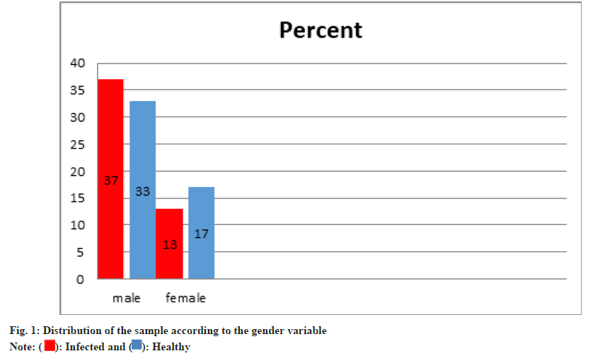
The data n Table 1 showed that the samples of the infected male reached (37), with a percentage of (74 %), while the number of infected females reached (13), with a percentage of (26 %). There are (33) healthy males, with a percentage of (66%), while the number of healthy females is (17) females, with a percentage of (34 %) (Table 1 and fig. 1).
| Sample | Gender | The number of sample | Percentage | Total |
|---|---|---|---|---|
| Infected | Male | 37 | 0.74 | 50 |
| Infected | Female | 13 | 0.26 | 50 |
| Healthy | Male | 33 | 0.66 | 50 |
| Healthy | Female | 17 | 0.34 | 50 |
Table 1: Distribution of the Sample by Gender
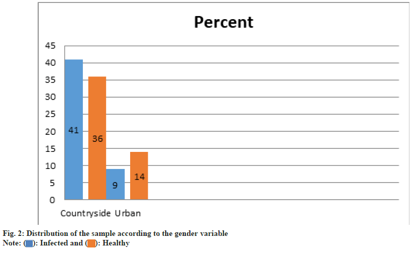
Through Table 2 it found that the samples of the infected countryside reached (41), with a percentage of (82 %), while the number of infected urban reached (9) with a percentage of (18 %). The number of healthy countryside residents there was (36) persons, with a percentage of (72 %), while the number of healthy urban is (14) persons. with a percentage of (28 %) (Table 2 and fig. 2).
| Sample | Residence | The number of sample | Percentage | Total |
|---|---|---|---|---|
| Infected | The countryside | 41 | 0.82 | 50 |
| Infected | Urban | 9 | 0.18 | 50 |
| Healthy | The countryside | 36 | 0.72 | 50 |
| Healthy | Urban | 14 | 0.28 | 50 |
Table 2: Distribution of the Sample by Residency
The study included measuring of biochemical parameters and measuring the following criteria: Determination of the level of urea in serum. Determination of the level of creatinine in the serum. After conducting a statistical analysis of the blood and biochemical parameters in patients with dengue fever, it was found that: Through the Table 3 and Table 4, we noted that there was no increased in the concentration of urea and creatinine as it was noted through the arithmetic mean of the samples of patients with dengue fever is closed to the arithmetic mean with the samples of healthy people, while the arithmetic mean between the two samples differed in the level of urea concentration, so the t-test was used to compare between the averages of the study population by testing the statistical hypotheses at the level of significance (p<0.05).
| Group | Infected group | Healthy group | ||
|---|---|---|---|---|
| Variables | Mean | Standard deviation | Mean | Standard |
| Creatinine | 1.32 | 1.6 | 1.06 | 0.21 |
| Urea | 28.78 | 13.58 | 37.54 | 14.95 |
Table 3: Comparison Between the Mean and Standard Deviation of Urea and Creatnine
| Axis | Type | Mean | Standard deviation | The computed (t) value | Degree of freedom | Indication level |
|---|---|---|---|---|---|---|
| Creatinine | Infected | 1.32 | 1.6 | 1.14 | 98 | 0.25 |
| Creatinine | Healthy | 1.06 | 0.21 | |||
| Urea | Infected | 28.79 | 13.58 | 3.06 | 98 | 0 |
| Urea | Healthy | 37.54 | 14.95 |
Table 4: Comparison Between the Mean, Standard Deviation, T-Value, Freedom Degree and Indication Level of Urea and Creatnine
As it is evident from Table 4 above, that the value of the significance level in creatinine between the samples of infected and healthy individuals was (0.25), which means that there are no statistically significant differences in the creatinine test. It was also showed that the value of the significance level in urea between the sample of infected and healthy subjects reached (0.00), which means that there are statistically significant differences between the samples of infected and healthy subjects in the level of urea.
The purpose of this study is to determine the effect of dengue fever on the kidney function test (creatinine and urea). In our study there were 100 sample tested a 50 of them infected with dengue fever other 50 samples were healthy persons. The researcher have used person correlation coefficient to study the effect of dengue fever on kidney function test which was noted that there was no effect on the concentration of urea and creatinine as it is noted through the arithmetic mean of the sample of dengue fever infected persons was closed to the arithmetic mean of sample of healthy person while the arithmetic mean between the two sample differed in level in urea concentration compared between the average of the study population by testing the so the test was used to statically hypothesis at the level of significant (p<0.05). Significant level in the creatinine between the sample of infected and healthy individual was (0.25), which means that there are no statistically significant difference in the creatnine test. It was also found that the value significant level in urea between the samples of infected and healthy subject researched (0.00), which means that there are statically significant difference between the sample of infected and healthy individual in the level of urea.
Our study disagreement with other study in which dengue is growing public health problem in India, and acute kidney infection is one of the least studied complication of dengue virus infection. Previous study reports suggested that DHF and DSS are associated with an increased risk of development acute kidney infection. This study have supported those finding among 90 patient with acute kidney infection, 66 (26-77 %) had sever dengue (DHF and DSS) and logistic regression demonstrated that patient with sever dengue develop acute kidney infection. Our study disagreement with other study done by (karlo j Lizarraga and Nayer) [32] which found that there are effect of dengue fever infection on kidney disease which can lead to kidney disease, Acute renal failure. Severe dengue infection, particularly DHF and DSS may give rise to multi-organ dysfunction. Acute Renal Failure (ARF) is a potential complication of severe dengue infection and typically associated with hypotension, rhabdomyolysis, or hemolysis. The prevalence of ARF was 1.6 % among 617 children with DHF in Colombia, 3.3 % in hospitalized adults with DHF, 4.9 % in 81 Chinese patients with DHF/DSS, and 5 % in DHF patients in Qatar[33]. The development of ARF in patients with dengue infection is associated with increased mortality. In Thailand, the prevalence of ARF in fatal DHF was 33.3 %, compared with 0.3 % in all DHF cases. The cause of disagreement with our study may be that the patients from whom we collected samples were receiving treatment for dengue infection, also all patient had no heart, diabetic, hypertension problems, so that may prevent the complication in kidney or prevent the impaired kidney functions.
At the end of the research found that the Dengue fever is a dangerous disease and the infection with DENV result in averring degrees of pathological conditions, ranging from mild asymptomatic dengue fever to severe DHF and DSS which may turn fatal. Should be take a careful from the symptoms from the degree of fever of dengue disease which is may be change to severe hemorrhagic and which the state on, fever and to become a fatal. The results from this research and from these tables found that there was no effect of dengue fever by general on kidney functions taken in consideration sample took from hospitalization patients and the patient had no heart, diabetic, hypertension problems so that may be the renal disorder did not appeared and the creatinine and urea level was normal.
Acknowledgments:
Princess Nourah bint Abdulrahman University Researchers Supporting Project number (PNURSP2025R891), Princess Nourah bint Abdulrahman University, Riyadh, Saudi Arabia.
Conflict of interests:
The authors declared no conflict of interests.
References
- Dengue and severe dengue Factsheet N°117". WHO. May 2015. Archived from the original on 2 September 2016. Retrieved 3 February 2016.
- Amarasinghe A, Kuritsky JN, Letson GW, Margolis HS. Dengue virus infection in Africa. Emerg Infect Dis 2011;17(8):1349.
- Murphy BR, Whitehead SS. Immune response to dengue virus and prospects for a vaccine. Ann Rev Immunol 2011;29(1):587-619.
[Crossref] [Google Scholar] [PubMed]
- Gubler DJ. Dengue/dengue haemorrhagic fever: History and current status. Novartis Found Symp 2006;277:3-16.
[Crossref] [Google Scholar] [PubMed]
- Kularatne SA. Dengue fever. BMJ 2015;351.
[Crossref] [Google Scholar] [PubMed]
- Anonymous. Dengue hemorrhagic fever, diagnosis, treatment and control. Geneva, Switzerland: World Health Organization; 1986.
- Gubler DJ. Dengue and dengue hemorrhagic fever. Clinical microbiology reviews. 1998;11(3):480-96.
[Crossref] [Google Scholar] [PubMed]
- Dietz V, Gubler DJ, Ortiz S, Kuno G, Casta-Velez A, Sather GE, et al. The 1986 dengue and dengue hemorrhagic fever epidemic in Puerto Rico: Epidemiologic and clinical observations. Puerto Rico Health Sci J 1996;15(3):201-10.
[Google Scholar] [PubMed]
- Patel ML, Himanshu D, Chaudhary SC, Atam V, Sachan R, Misra R, et al. Clinical characteristic and risk factors of acute kidney injury among dengue viral infections in adults: A retrospective analysis. Indian J Nephrol 2019;29(1):15-21.
[Google Scholar] [PubMed]
- Gould EA, Solomon T. Pathogenic flaviviruses. Lancet. 2008;371(9611):500-9.
[Crossref] [Google Scholar] [PubMed]
- Gubler DJ. Dengue. In: Monath TP, editor. Epidemiology ofarthropod-borne viral diseases. Boca Raton, Fla: CRC Press; 1988. p. 223-60.
- Westaway EG, Blok J. Taxonomy and evolutionary relationships of flaviviruses, In: Gubler DJ, Kuno G, editors. Dengue and dengue hemorrhagic fever. London, United Kingdom: CAB International; 1997. p. 147-73.
- Rico-Hesse R. Molecular evolution and distribution of dengue viruses type 1 and 2 in nature. Virology 1990;174(2):479-93.
[Crossref] [Google Scholar] [PubMed]
- Dengue: Guidelines for diagnosis, treatment, prevention and control. Geneva (Switzerland): World Health Organization: 2009. Chapter 1: Epidemiology, burden of disease and transmission; 2009. p. 3-21.
- Global strategy for dengue prevention and control 2012-2020. Geneva (Switzerland): World Health Organization; 2012. Par t3.3: Sustainable vector control; 2014. p. 14-6.
- Georgiev, Vassil. NationalInstitute of allergy and infectious diseases, NIH (1st edition). Totowa, N.J.: Humana; 2009. p. 268.
- Gubler DJ. Dengue viruses In Mahy BW, Van Regenmortel MH. Desk Encyclopedia of Humanand medical virology. Boston: Academic Press; 2010. p. 372-82.
- Wilder-Smith A, Chen LH, Massad E, Wilson ME. Threat of dengue to blood safety in dengue-endemic countries. Emer Infect Dis 2009;15(1):8.
[Crossref] [Google Scholar] [PubMed]
- Uhlén M, Fagerberg L, Hallström BM, Lindskog C, Oksvold P, Mardinoglu A, et al. Tissue-based map of the human proteome. Science 2015;347(6220):1260419.
[Crossref] [Google Scholar] [PubMed]
- Pocock Gillian; Richards, Christopher D. Human physiology: The basis of medicine (3rd ed.). Oxford: Oxford University Press; 2006. p. 349. J Tortora, Gerard. Principlesof anatomy and physiology Derrickson, Bryan. (12th ed.). Hoboken, NJ: John Wiley & Sons; 2010.
- Guyton and Hall, Medical Physiology. 11th edition. p. 314.
- Ganong. "Renal Function & Micturition". Review of Medical Physiology, 25th ed. McGraw-Hill Education; 2016. p. 677.
- Cooper R, Naclerio F, Allgrove J, Jimenez A. Creatine supplementation with specific view to exercise/sports performance: An update. J Int Soc Sports Nutr 2012;9(1):33.
[Crossref] [Google Scholar] [PubMed]
- Brosnan ME, Brosnan JT. The role of dietary creatine. Amino Acids 2016;48(8):1785-91.
[Crossref] [Google Scholar] [PubMed]
- Lewis, Sharon Mantik; Dirksen, Shannon Ruf); Heitkemper. Margaret M.; Bucher, Linda; Harding, Mariann. Medical-surgical nursing: Assessment and management of clinical problems (9th ed.). St. Louis, Missouri; 2013.
- What is a creatinine blood test? Low& high ranges Medicinenet; 2018.
- Shemesh O, Golbetz H, Kriss JP, Myers BD. Limitations of creatinine as a filtration marker in glomerulopathic patients. Kidney Int 1985;28(5):830-8.
[Crossref] [Google Scholar] [PubMed]
- Taylor E, Howard. Clinical chemistry. New York: John Wiley and Sons; 1989. p. 58-62.
- Gross JL, De Azevedo MJ, Silveiro SP, Canani LH, Caramori ML, Zelmanovitz T. Diabetic nephropathy: Diagnosis, prevention, and treatment. Diabetes Care 2005;28(1):164-76.
[Crossref] [Google Scholar] [PubMed]
- Isles CG, Paterson JR. Serum creatinine and urea: Make the most of these simple test. Br J Hosp Med 1996;55(8):513-6.
[Google Scholar] [PubMed]
- Nomenclatature of organic chemistry: IUPAC Recommendations and Perferred Names 2013 (blue book). Cambridge: 2014. p. 860-1.
- Lizarraga KJ, Nayer A. Dengue-associated kidney disease. J Nephropathol 2013;3(2):57.
- Méndez A, González G. Dengue haemorrhagic fever in children: Ten years of clinical experience. Biomedica 2003;23(2):180-93.
[Google Scholar] [PubMed]

 ): Infected and (
 ): Healthy
 ): Healthy


